Dog Skin Problems Pictures: 15 Conditions and When to See a Vet
Labeled pictures of 15 dog skin problems, from allergies and hot spots to mange, yeast, ringworm and lumps, plus a look-alike table, which ones spread to people and the signs that need a same-day vet visit.

BVMS MRCVS
Medically reviewed by Dr. Pippa Elliott, BVMS MRCVS · Last reviewed

This article contains affiliate links. Webvet may earn a commission when you buy through them, at no extra cost to you.
Most dog skin problems show up as one of six visible changes: redness, bumps, flakes or crusts, hair loss, darker skin, or open sores. The dog skin problems pictures here cover 15 conditions, grouped by what you see, with the key clue that separates each from its look-alikes and when it needs a vet.
Use dog skin problems pictures to narrow the list, not to reach a diagnosis from a photo alone. Many skin diseases look alike, and vets confirm the cause with tests such as skin scrapings, tape samples and fungal cultures, according to the Merck Veterinary Manual.
The WebVet page to read next depends on the condition: each section ends with its in-depth guide, including our picture guides to mange, yeast infections, hot spots, allergy rashes, staph infections, hives and black spots.
What does skin disease look like on a dog?
Skin disease on a dog looks like a change in the color, surface or coverage of the skin: pink or red patches, raised bumps, white or greasy flakes, thin or bald areas, darker skin, crusts or sores.
Healthy dog skin, by contrast, is smooth and clear, without grease, flakes or bumps (VCA Animal Hospitals). Match the change you see to the labeled photos that show it:
- 1Red, itchy skin, a rash or black specks in the fur: compare the allergic dermatitis, flea dirt and hot spot photos
- 2Raised bumps, pimples or welts: compare the pyoderma, hives, tick and wart photos
- 3Flakes, scale or crusts: compare the seborrhea, ringworm and mange photos
- 4Patchy hair loss or even thinning: compare the mange, ringworm and hormone-related hair loss photos
- 5Dark, thickened skin: compare the darkened skin and yeast photos
- 6Open sores, scabs or a crusted nose: compare the hot spot, lick granuloma and discoid lupus photos
- 7Skip the photos: go to an emergency vet now for facial swelling or breathing trouble, and see a vet today for pinpoint red spots or unexplained bruising
Under the closest photo, check the key clue to rule look-alikes in or out. If no photo fits, or two fit equally well, that is a question for your vet.
Early stage skin conditions can look like very little: an allergic dog often itches before the skin shows more than faint redness, and later hair loss, scabs and thick, dark skin come mostly from licking and scratching (Merck Veterinary Manual).
Check the skin every few days, including the armpits, groin and behind the ears, and watch for these early signs:
- Scratching, licking, chewing at the paws or rubbing the face
- Head shaking or ear scratching, since ear infections often come with skin allergy
- A musty smell, a common sign of a yeast infection
- Heavier shedding, a dry, dull coat or dandruff on the bedding
To photograph a skin problem for your vet:
- Use natural daylight by a window or outdoors, without flash glare.
- Part the fur with your fingers or a comb so the skin, not the coat, fills the frame.
- Take a close-up and a wider shot showing where on the body the change sits.
- Repeat every two to three days on the same spot, in the same light, so the dated photos track changes for your vet.
Types of dog skin diseases with pictures
Vets group the types of skin diseases in dogs by cause, and each type has a labeled photo below:
- Allergies: environmental, food, flea and contact allergy, plus hives
- Infections: bacteria (pyoderma), yeast and the ringworm fungus
- Parasites: mites, fleas and ticks
- Growths: warts, skin tags, cysts, fatty lumps and tumors
- Hormone disease: an underactive thyroid and Cushing's disease
- Immune disease: pemphigus and discoid lupus
- Other causes: hot spots, lick granulomas, seborrhea and darkened skin

A rigid cone for preventing licking. Measure for the correct size and check that your dog cannot reach the sore.
Webvet may earn a commission when you click through to Chewy, at no extra cost to you.
The table puts all 15 pictured conditions side by side. The key clue column names the sign that best separates look-alike conditions, the spreads to people column flags the few that can pass to your family, and the last column shows the usual vet treatment.
| Condition | What you see | Where it shows up | Key clue | Spreads to people? | Usual vet treatment |
|---|---|---|---|---|---|
| Allergic dermatitis | Red, itchy skin with reddish-brown saliva stains | Paws, face, ears, armpits, belly | Itch comes first, with no pus bumps early on | No | Itch control, flea control, food trial |
| Hot spot | One wet, red, oozing patch under matted fur | Head, legs, hips | Appears fast and hurts | No | Clipping, cleaning, itch control, a cone |
| Bacterial infection (pyoderma) | Red pimples with white centers, ring-shaped crusts | Belly and trunk | Pus bumps and peeling rings | Rarely | Medicated shampoo, antibiotics for deep infection |
| Yeast infection | Greasy, red, thickened, darkened skin | Skin folds, armpits, ears | Musty smell and brown greasy residue | No | Antifungal shampoo, oral antifungals if widespread |
| Ringworm | Round patches of broken hair and gray scale | Anywhere on the body | Round, dry and often not itchy | Yes, especially to children | Fungal culture, antifungal treatment for weeks |
| Mange | Crusts, red bumps and hair loss | Ears, elbows, hocks, belly; face and front legs in demodectic mange | Sarcoptic itches intensely; demodectic barely itches | Sarcoptic yes; demodectic no | Prescription mite medicine |
| Fleas and flea dirt | Black specks and small scabby bumps | Lower back, tail base, back of thighs | Specks turn red-brown on damp paper | Fleas bite people | Flea control for every pet |
| Ticks | Gray or brown bean-shaped bump | Head, neck, ears, shoulders | Legs at the skin | Ticks bite people too; dogs do not pass tick diseases directly | Removal, then watching for tick-borne illness |
| Hives | Raised round welts, puffy muzzle | Face, lips, ears, body | Appear within minutes; skin unbroken | No | Antihistamine and steroid |
| Lumps and growths | Warts, tags, cysts, fatty lumps or tumors | Anywhere; warts on lips and feet | Looks cannot rule out cancer | No | Needle sample or biopsy |
| Darkened skin | Flat, dark, velvety, thick skin | Groin, armpits, legs | Flat, not raised | No | Treating the underlying cause |
| Seborrhea | Dandruff flakes or greasy brown build-up | Back, face, flanks, folds | Flakes on calm skin; moving flakes are mites | No, unless mites | Medicated shampoo, treating the cause |
| Hormone-related hair loss | Even thinning, dull and dry coat | Both sides of the trunk, thighs, tail | Symmetrical and not itchy | No | Blood tests, hormone treatment |
| Lick granuloma and open sores | Raised, hairless, often wet sore | Front of the wrist or lower leg | Constant licking at one spot | No | Long antibiotic course, stopping the licking |
| Autoimmune crusting | Crusts, sores, a nose losing color | Nose, face, ears, footpads | Nose loses its cobblestone texture | No | Biopsy, immune-suppressing medicine |
Want a printable PDF of these types of dog skin diseases with pictures? There is no separate download: choose Save as PDF in your browser's print menu to keep the table and photos for your vet visit.
Red, itchy and inflamed skin
Red, itchy skin can look like a rash, a patch of dermatitis or scattered red spots. Common causes are allergies, parasites, hot spots and infections, which usually follow an allergy or parasite that has already damaged the skin (Merck Veterinary Manual). Red spots deserve a closer look:
- Small red, scabby bumps on the lower back and tail base usually mean flea bites or flea allergy.
- Red pimples with white centers point to bacterial infection.
- Tiny, flat, pinpoint red or purple spots on the belly, ears, gums or whites of the eyes are petechiae. Along with larger unexplained bruises, they are small bleeds linked to a low platelet count (VCA Animal Hospitals).
Pinpoint red spots need a vet the same day, even if your dog seems well. Low platelets can follow tick-borne infection, immune disease, drug reactions or cancer, and bleeding can also start inside the body.
If itching is the main problem and little shows yet, start with the common causes of itching.
Allergic dermatitis

Allergic skin is red and itchy, often with reddish-brown saliva staining where the dog licks (VCA Animal Hospitals), most often on the paws, face, ears, front-leg folds, armpits and belly (Merck Veterinary Manual).
Skin allergies come in four main types, and where the itch sits hints at which one:
- Environmental allergy (atopic dermatitis): paws, face, ears, armpits and belly, usually starting between 6 months and 3 years of age (Merck Veterinary Manual).
- Food allergy: the same itchy areas, but the itch stays about the same from season to season (Merck Veterinary Manual).
- Flea allergy: crusted, itchy bumps over the lower back, tail base and back of the thighs (Merck Veterinary Manual).
- Contact allergy: redness where the skin touches the trigger, such as the belly of a dog that lies on a treated lawn or carpet (Merck Veterinary Manual).
Over months, allergic skin can lose hair and turn thick, dark and leathery, and ear and skin infections can keep coming back (VCA Animal Hospitals).
Key clue: the itch comes first. Early allergic skin is red without pus-filled pimples or a single wet patch, which point to infection or a hot spot. Welts that rise within minutes are hives.
- Spreads to people: no. Allergies are the dog's own immune reaction, so they cannot pass to people or pets.
- See a vet: when the itch lasts more than a few days, or you see pimples, a smell or broken skin, which suggest infection has moved in.
For more allergic rash photos by body area, see our dog food allergy rash pictures, compare whether the trigger is food, fleas or environment, and if your vet suspects food, read how a hypoallergenic diet trial works.
Hot spots

A hot spot, which vets call pyotraumatic dermatitis or acute moist dermatitis, is a red, inflamed patch that appears quickly, oozes and may contain pus. It is often hidden under matted fur, most commonly on the head, legs or hips (VCA Animal Hospitals).
Hot spots run on a lick-itch cycle: licking irritates nerve endings, the spot itches more, and the dog licks harder (VCA Animal Hospitals). Environmental allergy, fleas, flea allergy and anal gland trouble usually start it, and the spot can grow fast and hurt (Virginia Tech Veterinary Teaching Hospital).
- Key clue: wet, sudden and painful. Ringworm, the classic look-alike, is dry, round, slow and usually not itchy. Our guide to telling ringworm or a hot spot shows both side by side.
- Spreads to people: no. The sore comes from the dog's own licking and scratching.
- See a vet: promptly, and the same day if it is spreading fast or very painful. A vet clips the hair, disinfects the skin with chlorhexidine, treats the itch and any infection, and may fit a cone to stop the licking (VCA Animal Hospitals).
For more photos by stage and body area, plus healing and prevention, see our hot spots on dogs pictures guide.
A Dogswell rigid recovery collar can help keep a hot spot out of reach. This listing is Small; measure your dog and choose a cone long enough to prevent licking.
What are the most common skin infections in dogs?
Bacterial skin infection, called pyoderma and usually caused by Staphylococcus pseudintermedius, is the skin infection vets treat most often: superficial pyoderma is the most frequent reason for antibiotic use in small animal practice (Merck Veterinary Manual).
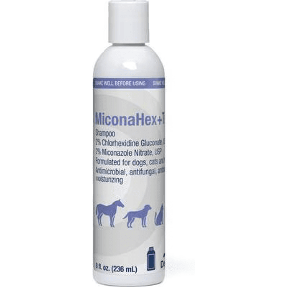
Miconazole and chlorhexidine shampoo for a veterinarian-directed bathing plan for confirmed skin infection.
Webvet may earn a commission when you click through to Chewy, at no extra cost to you.
Malassezia yeast infection is also common (Guillot and Bond, 2020). Ringworm is the fungal infection that matters most at home, because it spreads to people.
Most of these infections are secondary. They take hold because allergies, parasites, a skin injury or a hormone disease such as hypothyroidism or Cushing's disease has already weakened the skin, so the vet treats the infection and looks for the cause. Early stage dog skin infection photos tend to show only a few changes:
- A handful of small red bumps or pimples with white centers
- A ring of peeling skin where a pimple has burst
- A greasy, red patch in a skin fold or armpit
- A musty smell before much is visible
Bacterial skin infection (pyoderma)

Pyoderma looks like red, raised pimples, many with a white, pus-filled center, plus circular crusts, dry or flaky patches and hair loss (VCA Animal Hospitals). It shows up most often on the trunk and belly (Merck Veterinary Manual).
In short-coated dogs, including pit bull types, the first sign can be small tufts of hair standing up that look like hives, or a moth-eaten coat from patchy hair loss (VCA Animal Hospitals).
Deep pyoderma is more serious. Its hallmarks are pain, crusting, odor, and blood and pus oozing from the skin, and it needs a vet the same day (Merck Veterinary Manual).
- Key clue: pus-filled pimples and ring-shaped crusts. Flat allergic redness has neither.
- Spreads to people: rarely. Dog staph can occasionally infect people, so wash your hands after touching sores (Carroll and colleagues, PLOS Pathogens).
- Vet care: a swab or tape sample confirms the bacteria. Surface infections often clear with chlorhexidine or benzoyl peroxide shampoo alone, while deeper ones need antibiotics, usually for at least three to four weeks (VCA Animal Hospitals).
Our staph infection pictures guide shows superficial and deep pyoderma, puppy pyoderma and skin-fold infections in more detail.
What does a fungal infection look like on dog skin?
A fungal infection on dog skin usually looks like one of two things. Yeast (Malassezia) makes the skin red, greasy and itchy, with brown-black waxy material matting the hair and a musty smell, mostly in skin folds, armpits and ears (Guillot and Bond, 2020).
Ringworm, caused by fungi called dermatophytes, makes roughly round patches of hair loss with scale and broken hairs that are usually not itchy, and it can spread to people (VCA Animal Hospitals).
The two photos below show each one. Neither can be confirmed by looks alone: yeast is found on a tape or swab sample under the microscope, and ringworm is confirmed with a fungal culture or a PCR test.
Yeast infection (Malassezia dermatitis)

Early yeast infection shows itching, redness and a musty odor. With time the skin turns scaly and crusty, thickens into elephant skin and darkens (VCA Animal Hospitals), and greasy brown-black material mats the base of the hairs, especially in skin folds (Guillot and Bond, 2020).
Malassezia is a yeast that normally lives on dog skin and overgrows when something changes the skin, most often allergies, oily skin or a hormone disease (Guillot and Bond, 2020).
- Breeds at risk: West Highland White Terriers, Basset Hounds, Cocker Spaniels, Shih Tzus, Poodles, Dachshunds and several others (VCA Animal Hospitals).
- Key clue: grease and smell. Darkened skin with no greasy residue or odor is more likely simple hyperpigmentation.
- Spreads to people: no. Yeast dermatitis is not contagious.
- Treatment: a tape or swab sample confirms it. Antifungal and degreasing shampoos treat mild cases, oral antifungals are given for months in widespread cases, and the trigger has to be controlled (VCA Animal Hospitals).
If your vet recommends a miconazole-chlorhexidine wash for confirmed yeast, MiconaHex+Triz shampoo is one option. Follow the contact time and bathing schedule your vet gives you.
For more yeast infection pictures, from early redness to chronic black skin on the paws, belly and skin folds, see our dog skin yeast infection pictures.
Ringworm

Ringworm causes roughly circular patches of hair loss. As a patch grows, its center heals and hair may regrow, and the affected hairs are fragile and break easily. Patches are usually not itchy, though some become inflamed and scabby (VCA Animal Hospitals).
- Key clue: a round, dry, scaly patch with broken hairs that bothers the dog less than you would expect.
- Spreads to people: yes, especially to young children and people with weak immune systems, by direct contact or through shared bedding and brushes.
- Vet care: a fungal culture is the most accurate test. PCR is faster but can give false positives, and a Wood's lamp misses some cases. Treatment combines medicated dips, creams or shampoos with oral antifungal medicine for at least six weeks, plus daily cleaning at home (VCA Animal Hospitals).
With aggressive treatment, an infected dog stays contagious for about three weeks. See our ringworm on dogs pictures for more photos of new and healing patches.
Skin parasites: mites, fleas and ticks
Some skin parasites are easy to see and others are not. Fleas, flea dirt and ticks show up in pictures of parasites on dogs' skin, and chiggers appear as tiny orange-red dots that do not move, usually on the head, ears, feet or belly (Merck Veterinary Manual).
Mange mites live in or on the skin and need a skin scraping and a microscope to find. White flakes that seem to move along the back can be walking dandruff, caused by a mite called Cheyletiella that can also infest people.
Mange (sarcoptic and demodectic)

Sarcoptic mange, caused by the Sarcoptes scabiei mite, starts with sudden, intense itching and small, solid bumps that turn into thick, yellow crusts with hair loss. The ears, elbows, hocks, chest and belly are hit first (Merck Veterinary Manual).
Demodectic mange, caused by Demodex mites that live on every dog in small numbers, looks different: patchy hair loss around the eyes, lips and front legs, often with little itch, mostly in dogs under 12 to 18 months old (VCA Animal Hospitals).
A few small patches often clear on their own, but widespread demodectic mange, or mange that starts in an adult dog, needs a vet to look for an underlying disease (Merck Veterinary Manual).
- Key clue: sarcoptic mange itches intensely; demodectic mange usually does not.
- Spreads to people: sarcoptic mange is highly contagious to other dogs and causes itchy bumps on people until the mites die (VCA Animal Hospitals). Demodectic mange does not spread.
- Vet care: skin scrapings often miss sarcoptic mites, so vets may treat on suspicion. Both types respond to prescription isoxazoline parasite medicine, and every dog in the home is treated for sarcoptic mange.
Our mange in dogs pictures guide shows early and advanced stages of both types, along with walking dandruff and harvest mites.
Fleas and flea dirt

Flea dirt is flea droppings: reddish-black, comma-shaped specks in the fur, often at the base of the tail. Crush a few on a damp white paper towel. If they dissolve into reddish-brown smears, they are digested blood from fleas (Merck Veterinary Manual).
A fine-tooth flea comb pulled through the lower back and tail base catches fleas and flea dirt. Dogs with flea allergy get crusted, itchy bumps over the hips, tail base and thighs.
The fine teeth of the Safari Double Row Flea Comb help collect fleas and loose debris for the damp-paper test. Comb gently and avoid pulling over sore or broken skin.
- Spreads to people: fleas bite people and other pets too (Merck Veterinary Manual).
- See a vet: for a flea allergy rash or infected skin, and the same day for a weak, pale puppy, since heavy infestations can cause anemia in young puppies.
Year-round flea control means keeping every dog and cat in the home on a vet-recommended flea product all year, not only in summer, and treating the home during an outbreak. Clearing an infestation often takes up to three months (Merck Veterinary Manual).
Never put a dog flea product on a cat: some, including permethrin spot-ons, are toxic to cats, so ask your vet for a cat product.
See what fleas look like on dogs for flea photos and removal steps.
Ticks

A tick that has just attached looks flat. After feeding for a few days it turns rounded, gray or brown and bean-shaped, with small legs at the skin (Merck Veterinary Manual). Ticks favor the head, neck, shoulders, ears and the area under the tail.
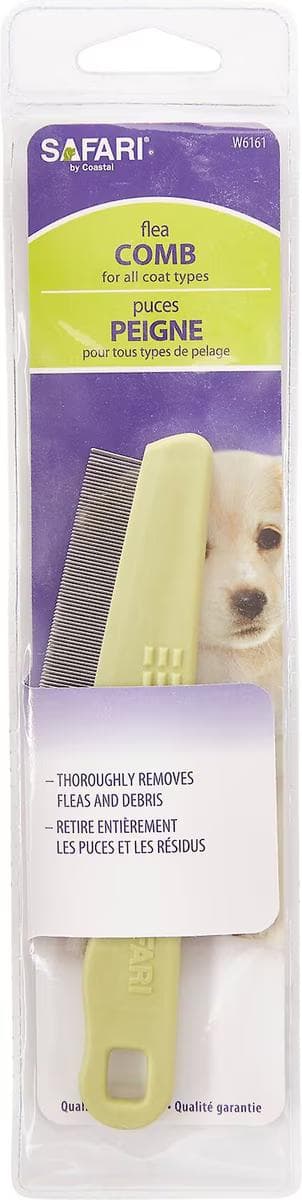
A fine-tooth comb for collecting fleas and flea dirt while checking the coat.
Webvet may earn a commission when you click through to Chewy, at no extra cost to you.
- Key clue: legs at the skin and a body that grows as it feeds. A skin tag hangs on a stalk and a wart has a cauliflower surface, and neither has legs.
- Remove it promptly: grasp the tick with fine tweezers close to the skin and pull gently. Never use bare hands or a hot match.
- See a vet: if the bite site turns red or swells, or your dog seems unwell in the weeks after, because ticks spread diseases such as Lyme disease, ehrlichiosis and Rocky Mountain spotted fever.
See what tick bites look like for more photos of bites and bite reactions.
 $65 · no membership
Is your dog's itching or skin issue getting worse?
A licensed vet can examine your dog over video and prescribe treatment online when it's appropriate.
 Talk to a vet
- Licensed vets
- All 50 states
- same-day Rx

Webvet may earn a commission if you book.
Bumps on dog skin: pimples, welts and lumps
Bumps on a dog's skin sort into a few groups by how they look and how fast they appeared. Find the closest match, then read the matching section:
- Red pimples with white centers: bacterial infection.
- Tiny red, scabby bumps on the lower back and tail base: flea bites or flea allergy.
- Raised tufts of hair on a short-coated dog such as a pit bull type: often a hair-follicle infection that can mimic hives.
- Itchy welts that appear within minutes: hives from an allergic reaction.
- A white bump: usually a pustule, or a cyst that may leak cheesy gray, brown or yellowish material (Merck Veterinary Manual).
- A gray, bean-shaped bump with legs: a tick.
- Cauliflower-like bumps on the lips or feet of a young dog: likely warts.
- A firm lump, or one that grows or changes: a growth that needs a needle sample.
Treatment follows the cause, so the picture only tells you where to start. Pimples need a vet swab and usually a medicated shampoo, hives need vet-directed anti-allergy care, and lumps need a fine-needle sample, because nobody can tell a harmless lump from cancer by looking.
Hives and insect bites

Hives, or urticaria, are raised, round, itchy welts that lift the hair, from about 5 mm to several centimeters across, often on the face, lips and ears (VCA Animal Hospitals). The muzzle and eyelids can puff up, and hives usually appear within 20 minutes of contact with the trigger (Merck Veterinary Manual).
Common triggers include insect bites and stings, vaccines, medicines, foods and pollen. Mild hives often fade on their own within 12 to 48 hours, but vets usually treat them because it is hard to predict whether they will settle or get worse (Merck Veterinary Manual).
- Key clue: the welts appear fast and the skin stays unbroken, with no pus or crusts.
- Spreads to people: no.
- Vet care: an antihistamine and a steroid, often by injection, can shrink hives, sometimes within minutes (VCA Animal Hospitals). Give an antihistamine at home only at a dose your vet confirms for your dog; our Benadryl dose guidance explains the details.
See pictures of hives on dogs for more welts, facial swelling and bite reactions.
Lumps and growths

Viral warts, or papillomas, are small, pale, cauliflower-like growths with fingerlike projections, most common on the lips and mouth of young dogs (Merck Veterinary Manual).
Other lumps are harder to call. Telling a harmless lump from cancer takes a lab, so a vet takes a fine-needle sample of cells or a small biopsy (Merck Veterinary Manual). Common lumps and where to read more:
- Warts: mostly in young dogs, on the lips, mouth or feet. Many go away within one to two months, and they pass between dogs but not to people (VCA Animal Hospitals). More photos: dog warts.
- Skin tags: soft, stalk-like growths on older dogs, often with a wart-like surface. See skin tags on dogs.
- Cysts: round lumps that may leak gray, brown or yellowish cheesy material. See dog cysts.
- Histiocytomas: smooth, pink, raised, hairless button lumps, usually in dogs under about 3.5 years, and most disappear within 2 to 3 months (Merck Veterinary Manual). See histiocytoma.
- Lipomas: soft, movable lumps of fat under the skin, common in older, overweight dogs. See fatty lumps (lipomas).
- Mast cell tumors: can look like an insect bite, a wart or an allergic swelling, and may change size from day to day (VCA Animal Hospitals). See mast cell tumor pictures.
Not sure which one you have? Start with our guide to lumps on dogs, and book a vet visit for any new lump, sooner if it grows quickly, bleeds or changes.
Black spots and darkened skin

Darkened skin, or hyperpigmentation, is light brown to black, velvety, thickened and often hairless, usually on the legs and groin first (Merck Veterinary Manual). It lies flat against the body.
In Dachshunds, a primary form shows by about 1 year of age and can be managed but not cured.
In other dogs the darkening is usually secondary: the skin's response to long-term inflammation or friction from allergies, skin infections, hormone disease, obesity or contact irritation. It fades once the cause is treated. Other dark changes mean something different:
- Black specks in the fur: likely flea dirt, so do the damp paper towel test.
- Dark, greasy, smelly skin in folds: points to yeast.
- Blackheads on a thin-skinned belly with hair loss: can be a sign of Cushing's disease, covered under hormone disease below.
- A raised, growing, bleeding or ulcerated dark spot: needs a vet to rule out melanoma, which usually appears as a raised, often ulcerated lump that may or may not be dark (Merck Veterinary Manual).
Flat darkened skin does not spread to people or pets. See black spots on dog skin pictures for normal pigment spots, blackheads and when a dark spot is a concern.
Flaky skin and hair loss
Flakes and hair loss sort mainly by itch. Itchy flakes or bald patches point to parasites, allergy or infection. Even, non-itchy thinning on both sides of the body points to hormone disease, which vets confirm with blood tests (Merck Veterinary Manual).
Scaly skin also appears with ringworm, mange and autoimmune disease, so check the whole dog. The pattern of hair loss narrows it further:
- Round, scaly patches with broken hairs: ringworm
- Patches around the eyes, lips and front legs of a young dog: demodectic mange
- Thinning over the lower back, tail base and thighs: flea allergy
- Even thinning on both sides of the trunk that spares the head and legs: hormone disease

A 2.5% benzoyl peroxide shampoo for vet-directed care of greasy, scaly skin.
Webvet may earn a commission when you click through to Chewy, at no extra cost to you.
Dry, flaky or greasy skin (seborrhea)

Seborrhea shows up as dry flakes like dandruff, often seen on the dog's bedding, or as a greasy, brown, waxy build-up on the skin, often with a smell. It mostly affects the back, face and flanks and is worse in the skin folds of the feet, neck, lips, armpits and belly (VCA Animal Hospitals).
Primary seborrhea is inherited in breeds such as Cocker Spaniels, West Highland White Terriers and Basset Hounds. Secondary seborrhea is more common and follows allergies, parasites, hormone problems such as thyroid or Cushing's disease, or fungal and bacterial infections (VCA Animal Hospitals).
The oily form, which vets call seborrhea oleosa, often travels with yeast, because extra skin oil helps Malassezia overgrow (VCA Animal Hospitals).
- Key clue: flakes sitting on calm, pale skin suggest seborrhea or dry skin. Flakes that move are walking dandruff mites, which spread to other pets and people.
- Watch for: red, scaly skin in a middle-aged or older dog that does not improve with allergy treatment. Early cutaneous lymphoma, a skin cancer, can be confused with allergic, hormonal or infectious skin disease (Merck Veterinary Manual).
- Vet care: anti-seborrheic shampoos, mousses or sprays and omega-3 fatty acid supplements, with the main aim of treating the underlying cause.
Our dandruff and flaky skin checker helps you sort flaky skin by the other signs your dog has.
Hair loss and skin changes from hormone disease

Hypothyroidism, an underactive thyroid, causes non-itchy hair thinning or baldness that usually matches on both sides of the body, on the sides and underside of the trunk, the back of the thighs, the tail, the neck and the top of the nose (Merck Veterinary Manual).
The coat turns dry and dull and sheds heavily, and clipped hair is slow to grow back. Hypothyroidism is most common in dogs 4 to 10 years old, usually mid- to large-size breeds such as Golden Retrievers and Doberman Pinschers. Other signs of hypothyroidism include:
- Weight gain without a bigger appetite
- Tiredness and feeling the cold
- Darker skin
- Skin and ear infections that keep coming back (VCA Animal Hospitals)
Cushing's disease, an excess of the hormone cortisol, mostly affects dogs 7 to 12 years old (Merck Veterinary Manual). Its signs include:
- Even hair loss on the trunk that spares the head and feet
- Thin, fragile skin with blackheads (comedones) and easy bruising
- Darker skin and calcium deposits in the skin (calcinosis cutis)
- A pot belly, panting, a bigger appetite, and more drinking and urinating
Diabetes can mark the skin too. Older dogs with diabetes or liver disease can develop necrolytic dermatitis, with red, crusted, oozing skin on the footpads, face and lower legs (Merck Veterinary Manual), and demodectic mange that starts in adulthood is linked to diabetes, Cushing's disease and hypothyroidism.
Repeated infections are a clue in their own right. A middle-aged dog whose skin or ear infections keep coming back may need thyroid or adrenal testing, because both diseases leave the skin open to infection (Merck Veterinary Manual).
Hormone diseases are confirmed with blood tests, not by looks. Hypothyroidism is treated with daily thyroid hormone for life, and the coat can take up to three months to improve. Our hair loss symptom checker sorts hair loss by your dog's other signs.
Open sores, ulcers and scabs

A lick granuloma, also called acral lick dermatitis, is a raised, thickened, hairless sore that starts red and crusted and can erode into an open ulcer (Clinician's Brief). It usually forms on the front of the wrist or lower foreleg of a dog that licks the same spot over and over (VCA Animal Hospitals).
Lick granulomas mostly affect medium to large breeds, especially Doberman Pinschers, Great Danes, Labradors, Weimaraners, Golden Retrievers and Boxers (VCA Animal Hospitals). Allergy is the leading cause, and deep infection often sits under the surface, so vets treat a lick granuloma as allergic skin disease until proven otherwise (Clinician's Brief).
Joint pain, anxiety and compulsive licking can also drive it. Treatment usually combines a long antibiotic course and pain relief with a cone or bandage, plus more exercise and play to break the habit (VCA Animal Hospitals). Other common causes of open sores and scabs:
- Deep bacterial infection: pain, crusting, odor, and blood and pus.
- Hot spots: a wet, oozing sore that appeared quickly.
- Scratching wounds: scabs from mange, fleas or allergies, which clear only when the itch is treated.
- Skin tumors: squamous cell carcinoma often looks like a firm, raised, ulcerated patch (Merck Veterinary Manual).
- Autoimmune disease: crusts and sores on the face, ears and feet, described below.
Open sores, ulcers and deep scabs need a vet. Go the same day if a sore is deep, bleeding, smelly or very painful, and book a visit for any sore that is not clearly healing within about a week.
Our wounds and bleeding checker helps you judge how urgent a sore is.
Crusts and sores from autoimmune disease

In discoid lupus, the nose loses its black color and its bumpy, cobblestone texture and turns smooth, with sores, ulcers and crusts at the edges. The lips, the skin around the eyes and the ear flaps can be affected too (VCA Animal Hospitals).
Autoimmune skin disease happens when the immune system attacks the skin. Sunlight makes discoid lupus worse, and German Shepherds, Siberian Huskies, Collies and Shetland Sheepdogs are prone to it (Cornell University College of Veterinary Medicine).
Pemphigus foliaceus is the most common autoimmune skin disease in dogs. Pustules break open into crusts and scabs around the eyes, ears, footpads, groin and bridge of the nose (VCA Animal Hospitals), and without treatment the crusting can spread from the face and ears to the feet and trunk (VCA Animal Hospitals).
- Key clue: pemphigus usually crusts both ears and the face at the same time, while sarcoptic mange, which also crusts the ears, itches intensely.
- Triggers: some cases follow a drug reaction or strong sun exposure.
- Breeds at risk: Akitas and Chow Chows especially, plus Cocker Spaniels, Labrador Retrievers, Dachshunds and English Bulldogs.
- Warning signs: fever, poor appetite or low energy alongside the crusting, which can happen in severe cases.
- Diagnosis and care: both diseases need a skin biopsy. Treatment suppresses the immune system with steroids or other drugs, often for months or for life, and dogs with autoimmune skin disease should avoid strong sun.
- Spreads to people: no.
Which breeds of dogs are most prone to skin problems?
Breeds prone to skin problems include Boxers, Labrador and Golden Retrievers, West Highland White Terriers, Shih Tzus, Cocker Spaniels, Bulldogs and other flat-faced breeds, German Shepherds, Dachshunds and Shar-Peis. Each breed is prone to particular conditions, listed here by condition.
To check your dog, find the condition you suspect below. If your breed is named next to it, your dog is at higher risk for that problem, though not certain to get it.
- Allergies (atopic dermatitis): Boston Terriers, Boxers, Shar-Peis, Dalmatians, Golden and Labrador Retrievers, Lhasa Apsos, Scottish Terriers, Shih Tzus, West Highland White Terriers and Wire Fox Terriers (Merck Veterinary Manual).
- Yeast infection: West Highland White Terriers, Basset Hounds, Cocker Spaniels, Shih Tzus, Poodles, Dachshunds and Boxers (VCA Animal Hospitals).
- Skin-fold infection: English Bulldogs, French Bulldogs and Pugs had by far the highest odds in a large UK study of veterinary records (O'Neill and colleagues, 2022).
- Deep, recurring bacterial infection: German Shepherds.
- Demodectic mange: it can run in families, so vets advise against breeding affected dogs (Merck Veterinary Manual).
- Pemphigus foliaceus: Akitas and Chow Chows, plus Cocker Spaniels, Labradors, Dachshunds and English Bulldogs.
- Discoid lupus: German Shepherds, Siberian Huskies, Collies and Shetland Sheepdogs.
- Lick granuloma: Doberman Pinschers, Great Danes, Labradors, Weimaraners, Golden Retrievers and Boxers.
- Primary seborrhea: Cocker Spaniels, West Highland White Terriers and Basset Hounds.
- Primary darkened skin: Dachshunds.
- Hypothyroidism: Golden Retrievers, Doberman Pinschers, Irish Setters, Miniature Schnauzers, Dachshunds, Cocker Spaniels and Airedale Terriers.
- Cushing's disease: Poodles, especially Miniature Poodles, Dachshunds, Boxers, Boston Terriers, Yorkshire Terriers and Staffordshire terriers (Merck Veterinary Manual).
- Mast cell tumors: Boxers, English Bulldogs, Boston Terriers, Pugs, Golden and Labrador Retrievers, Beagles, Shar-Peis and several other breeds.
- Ringworm: lifestyle matters more than breed; young dogs, dogs that roam or hunt, and dogs in warm environments are at higher risk (Merck Veterinary Manual).
- Fleas, ticks and insect bites: any exposed dog can be affected, whatever its breed.
Five popular breeds in more detail:
- Boxers appear on the allergy, yeast and lick granuloma lists and are also prone to mast cell tumors, so any new lump on a Boxer deserves a needle sample (VCA Animal Hospitals).
- Labrador Retrievers are prone to allergies, lick granulomas and pemphigus, so itchy paws and a licked foreleg deserve an early look.
- Golden Retrievers share the allergy and lick granuloma risk and are a classic hypothyroid breed, so even, non-itchy thinning in a middle-aged Golden is worth a thyroid test.
- Bulldogs, English and French, carry the highest skin-fold risk, and English Bulldogs are also prone to pemphigus and mast cell tumors.
- German Shepherds can develop a deep, recurring bacterial infection called German Shepherd dog pyoderma, tied to allergies, hypothyroidism or a weak immune response, and they are prone to discoid lupus (Rosser, 1997).
A prone breed is not a diagnosis, and a dog whose breed is not listed, including a mixed breed, can still get any of these conditions. Breed risk tells you which photos to compare first and which tests your vet may suggest, so mention your dog's breed or likely mix at the visit.
When to see a vet about a skin problem
Most skin problems need a vet visit sooner or later, because the cause has to be confirmed before treatment works. These same-day vet red flags come first:
Book a routine visit within a week for:
- Itching, licking or chewing that lasts more than a few days
- Ear or skin infections that keep coming back
- Hair loss that is spreading, even without itch
- Any new lump or bump
- Flakes, dark patches or a thin coat that does not improve
Watching at home with dated photos is reasonable only for a small change that is not spreading, not itchy and not painful, in a dog that is eating, playing and acting normally. If it grows, spreads or starts to bother your dog, book the visit.
Bring the dated photos and a list of the flea products, food and any creams you have used.
At the visit, the vet asks when and where the problem started, then chooses tests based on the look (Merck Veterinary Manual):
- Flea combing to collect fleas, flea dirt and skin debris
- Skin scraping to look for mites under the microscope
- Tape or swab cytology to find bacteria and yeast
- Fungal culture for suspected ringworm
- Blood tests for thyroid and adrenal disease
- A biopsy when the signs are severe or unusual, or do not respond to treatment
- A food trial or allergy testing when the pattern points to allergy
How to cure a dog with skin problems?
Whether a skin problem can be cured depends on the cause. Parasites and most infections can usually be cleared, while allergies, hormone diseases and autoimmune diseases are controlled long term rather than cured (Merck Veterinary Manual; VCA Animal Hospitals).
Curing a skin problem takes three steps:
- Find the cause with a vet exam and tests first. Guesswork often fails, and bathing before the visit can remove the crusts a vet needs to see (Merck Veterinary Manual).
- Treat that cause and any infection on top of it, as shown for each type below.
- Keep up prevention, such as year-round flea control, so it does not come back.
Treatment for each type usually looks like this:
- Parasites: prescription parasite medicine for every dog in contact, and flea control for every pet in the home. Clearing fleas from a house can take up to three months, and widespread demodectic mange is treated for several months, until two monthly skin scrapings in a row are clear.
- Bacterial infection: chlorhexidine or benzoyl peroxide shampoo, which is often enough for surface infections, and antibiotics for deeper ones, continued until the lesions and skin samples show the infection has cleared (Merck Veterinary Manual), which VCA notes usually takes at least three to four weeks (VCA Animal Hospitals).
- Yeast: antifungal and degreasing shampoo, with oral antifungal medicine for widespread cases, often for months.
- Ringworm: topical and oral antifungal medicine for at least six weeks, plus daily cleaning at home.
- Allergies: itch control with oclacitinib, lokivetmab, short steroid courses or cyclosporine; allergen immunotherapy, which Merck notes remains most veterinary dermatologists' treatment of choice for environmental allergy; strict flea control; and a food trial of up to three months when food is suspected.
- Hormone disease: daily thyroid hormone for hypothyroidism, with the coat improving over up to three months, or medicine that lowers cortisol for Cushing's disease.
- Autoimmune disease: immune-suppressing medicine, often long term (VCA Animal Hospitals).
Most skin treatment takes weeks, not days. Finish the full course even when the skin looks better: Merck advises antibiotics until the skin fully heals and mange treatment until scrapings are clear.
Safe home care while you wait for the vet should keep the skin from getting worse without hiding the problem:
- Keep every dog and cat on year-round flea control.
- Use a cone to stop licking and chewing at a sore.
- Wash bedding, and wash your hands after handling your dog, if ringworm or mange is possible.
- Keep taking dated photos so your vet can see how the skin has changed.
- Avoid human medicines, creams and painkillers unless your vet says otherwise. Merck warns that human anti-inflammatory painkillers should not be given to pets without veterinary direction, and dogs can lick creams off their skin and swallow them (Merck Veterinary Manual).
Bathe only with a shampoo made for dogs, because human shampoo, even baby shampoo, is too harsh for dog skin (VCA Animal Hospitals). Our guide to soothing or medicated dog shampoo covers what to look for, but ask your vet before bathing broken or oozing skin.
More questions about dog skin problems
Frequently Asked Questions
Can you tell what a dog skin problem is from a picture?
Not reliably. A picture can narrow the list, but look-alike conditions are common: a hot spot, ringworm and early mange can all start as a red or bald patch.
Vets confirm the cause with tests such as a skin scraping for mites, cytology for bacteria and yeast, a fungal culture for ringworm or a biopsy.
No photo alone can give a diagnosis, but dated photos that show how the skin changed over several days are worth bringing to the appointment.
Which dog skin problems are contagious to people?
Only a few skin problems spread to people. Ringworm spreads easily, especially to young children and people with weak immune systems, and sarcoptic mange causes intensely itchy bumps on people until the mites die.
Walking dandruff mites can infest people as well, and fleas and ticks bite people too. Bacterial skin infections (pyoderma) rarely spread to people. Allergies, hot spots, yeast infections and demodectic mange do not spread.
If contagion is possible, wash your hands after handling your dog, wash the bedding and see your own doctor for any itchy rash. Read more on whether ringworm is contagious to humans.
What is the best thing to give a dog with skin allergies?
The best treatment depends on the allergy type, so the first step is working out which one your dog has. Flea allergy calls for strict flea control on every pet in the home. Food allergy calls for a vet-guided elimination diet for up to three months.
Environmental allergy usually needs prescription anti-itch medicine such as oclacitinib (Apoquel) or lokivetmab (Cytopoint), and often allergy shots. Merck notes that antihistamines do little during a flare, though they can help alongside other treatment.
What can I give my dog for allergies without going to the vet?
Without a vet, stick to steps that are safe for any itchy dog. Keep every pet on flea prevention, because even a few flea bites add to the itch. Bathe your dog with a gentle shampoo made for dogs, since human shampoo is too harsh for dog skin.
Fit a cone if your dog is licking a sore raw. Do not give human medicines, including painkillers or antihistamines, unless your vet confirms the drug and the dose, and book a visit if the itch lasts more than a few days.
What is the best anti-itch supplement for dogs?
Omega-3 fatty acids, such as those in fish oil, are the supplement veterinary references point to for itchy or flaky skin, but only as an add-on to other treatment.
Merck notes that essential fatty acids can take up to two months to show an effect and work best alongside other allergy care, and VCA lists omega-3 supplements as part of seborrhea treatment. Ask your vet for a product and amount that suits your dog, and see our guide to omega-3 fatty acids for dogs.
$65 · no membershipIs your dog's itching or skin issue getting worse?
A licensed vet can examine your dog over video and prescribe treatment online when it's appropriate.
Talk to a vet- Licensed vets
- All 50 states
- same-day Rx
Webvet may earn a commission if you book.

Editor
The Webvet Editorial Team is the in-house group of pet-care editors and writers behind Webvet, operated by Smart Pet Collective. The team researches, writes, and maintains Webvet's pet health, behavior, and medication content. Every article follows a defined editorial process: research from reputable veterinary and scientific sources, careful drafting, mandatory review of medical content by a credentialed veterinarian, and dated publication. Health and medication articles are medically reviewed by a licensed veterinary professional before they go live and are kept current over time.
Veterinarian · BVMS MRCVS
Dr. Pippa Elliott, BVMS, MRCVS, is a veterinarian with nearly 30 years of experience in companion animal practice. Dr. Elliott earned her Bachelor of Veterinary Medicine and Surgery from the University of Glasgow. She was also designated a Member of the Royal College of Veterinary Surgeons. Married with 2 grown-up kids, Dr. Elliott has a naughty Puggle named Poggle, 3 cats and a bearded dragon.
Related reading

Pictures of Black Spots on Dog Skin and What Each One Means

Skin Rash Dog Food Allergy Pictures: What Allergic Skin Looks Like

Dog Skin Yeast Infection Pictures: Paws, Belly, Skin Folds and Face
