Lump on Dog: A Vet's Guide to When to Worry
Most lumps on dogs are benign, but only testing can confirm it. Run your dog's lump through our 60-second triage table, match it against photos of the 8 common types, and learn exactly which warning signs mean a same-day vet visit.

BVMS MRCVS
Medically reviewed by Dr. Pippa Elliott, BVMS MRCVS · Last reviewed

This article contains affiliate links. Webvet may earn a commission when you buy through them, at no extra cost to you.
Finding a lump on dog skin while you pet or groom your dog is one of the most common reasons owners call a veterinarian, and one of the scariest moments in pet ownership. Here is the honest, evidence-based answer up front: most lumps on dogs are benign. Fatty lipomas, cysts, warts, and skin tags vastly outnumber cancerous masses, especially in young and middle-aged dogs. But a small percentage of lumps are malignant, and no one, including your veterinarian, can reliably tell benign from cancerous by look and feel alone.
That is why this guide is built around triage, not diagnosis. In the next few minutes you will run your dog's lump through a 60-second triage table, compare it against a labeled photo ID chart of the 8 common lump types, and learn exactly which warning signs mean a same-day vet visit rather than watchful waiting.
- 1Most lumps on dogs are benign, but only a vet test (fine needle aspiration or biopsy) can confirm what a lump actually is.
- 2Use the 60-second triage table below: feel, mobility, growth speed, and skin changes map your dog's lump to a risk tier.
- 3Rapid growth, bleeding, ulceration, pain, or a hard lump fixed to underlying tissue all warrant a prompt vet visit.
- 4Any new lump that persists longer than a month, or any lump that changes, deserves a vet exam even if it seems harmless.
- 5Never squeeze, pop, or attempt to remove a lump at home.

 Sponsored
Want a vet's take on your dog's skin or coat?
Talk to a licensed vet at Dutch over video, often the same day. They can recommend treatment and prescribe it when it's appropriate.
Use code Pets30
Plans start at $11/month · covers up to 5 pets
- Licensed vets in most states
- Rx for 150+ conditions
- 4.7 on Trustpilot
The 60-Second Lump Triage Table
Should you be concerned if you find a lump on your dog? Concern should scale with what the lump is doing, not just the fact that it exists. Veterinary oncologists consistently point to the same observable criteria: how the lump feels, whether it moves freely under the skin, how fast it is growing, and whether the overlying skin is changing. The table below maps those four criteria to a risk tier and a concrete next step, so you can decide in about 60 seconds whether you are booking a routine appointment or calling for a same-day vet visit.
| What you observe | Typical examples | Risk tier | Next step |
|---|---|---|---|
| Soft, freely movable under the skin, slow or no growth, normal skin over it | Lipoma, sebaceous cyst | Lower risk | Book a routine vet exam; measure and photo-log the lump now |
| Small, wart-like, pink or pigmented, stable for weeks | Papilloma (wart), skin tag | Lower risk | Routine vet exam; monitor monthly for change |
| Firm but movable, appeared over days to weeks, no skin changes | Histiocytoma, early cyst | Moderate risk | Vet exam within 1-2 weeks; fine needle aspiration likely |
| Red, raised, itchy, or changing size day to day | Possible mast cell tumor, insect bite reaction | Higher risk | Vet visit within days; do not squeeze or rub |
| Hard, fixed in place, growing quickly, or attached to underlying tissue | Possible sarcoma or other malignancy | Higher risk | Prompt vet visit; ask about aspiration and biopsy |
| Bleeding, ulcerated, painful, or interfering with breathing, eating, or walking | Ulcerated mass, abscess, aggressive tumor | Urgent | Same-day vet care |
| Swelling that appeared overnight, warm or painful to touch | Abscess, hematoma | Urgent | Same-day vet call; these often need drainage and antibiotics |

Sponsored Lemonade pet insurance link. Webvet may earn a commission.
The when-to-worry criteria usually get buried in long paragraphs, so this risk-tier table structures the decision at a glance. One row deserves emphasis: a hard immovable lump on dog skin, one that feels fixed to the muscle or tissue beneath rather than sliding under your fingers, sits in the higher-risk tier no matter how slowly it grew.
Two important caveats. First, risk tier is about how quickly to see a vet, not a diagnosis. A soft movable lump can occasionally be something serious, which is why even lower-risk lumps earn a vet exam rather than a shrug. Second, growth speed matters more than size. A pea-sized lump that doubled in two weeks is more concerning than a golf-ball lipoma that has not changed in two years.
The Merck Veterinary Manual notes that skin tumors are among the most frequently diagnosed tumors in dogs, in part because they are the ones owners can actually see and feel (Merck Veterinary Manual, Tumors of the Skin in Dogs). Early detection is a genuine advantage: small masses caught early are simpler and cheaper to test and, when they do turn out to be malignant, far easier to remove completely.
Photo ID Chart: The 8 Common Types of Lumps on Dogs
This photo ID chart works as a labeled photo ID grid: each lump type gets its own labeled photograph plus a link to that condition's full guide. It covers the 8 common lump types veterinarians see most often. Compare your dog's lump against each photo, then follow the link under the closest match to that condition's full guide. Remember that visual matching narrows the possibilities; it never replaces a fine needle aspiration or biopsy.
1. Lipoma (Soft Fatty Lump)

A lipoma is a benign tumor of fat cells and the single most common soft lump in middle-aged and older dogs. It feels soft to slightly rubbery, sits just under the skin, and typically moves freely when you push it. Lipomas usually grow slowly over months to years and rarely bother the dog unless they get large or grow in an awkward spot like the armpit. Read the full guide to lipomas in dogs for removal criteria, costs, and the rare infiltrative form.
2. Sebaceous Cyst

A sebaceous cyst is a blocked oil gland that fills with thick, pasty material. It feels firmer than a lipoma, often round and well-defined, and sometimes shows a small dark pore at its center. Cysts can rupture, drain, and refill, and squeezing one drives material into surrounding tissue and invites infection. Our guide to cysts on dogs covers the different cyst types and when removal makes sense.
3. Mast Cell Tumor

A mast cell tumor is the most common malignant skin tumor in dogs, and it is a shape-shifter: it can look like a red raised bump, a soft lump, or even mimic a lipoma. The classic tell is change, swelling up and shrinking down over days, or getting red and itchy after being handled. Because handling can trigger histamine release, never rub or squeeze a suspicious red lump. See the full guide to mast cell tumors in dogs, and browse mast cell tumor pictures to compare more examples.
4. Histiocytoma (Button Tumor)

A histiocytoma is a benign immune-cell tumor that shows up mostly in dogs under about 3 years old. It looks like a small, pink to red, hairless dome, often on the head, ears, or legs, and it appears fast, sometimes within a week or two. The good news: most histiocytomas regress on their own within a few months. The full guide to histiocytomas in dogs explains how vets confirm one and when removal is warranted.

A collar-mounted sensor and app for activity, rest, sleep, respiratory rate, heart-rate trends, itch behaviors, and more.
- Builds a personalized baseline over time
- Tracks trends between veterinary visits
- Generates a month-long shareable report
Health monitoring for dogs and cats
Webvet may earn a commission when you click through to Maven Pet, at no extra cost to you.
5. Wart (Papilloma)

Dog warts, caused by the canine papillomavirus, have a distinctive cauliflower texture. In young dogs they cluster around the mouth, lips, and eyes; in seniors, single warts can pop up anywhere. Most viral warts in young dogs resolve on their own as immunity develops. Our guide to warts on dogs covers the contagious oral form, senior warts, and treatment options.
6. Skin Tag

A skin tag is a soft, floppy outgrowth of skin, often on a narrow stalk. Skin tags are benign, common in older dogs, and usually only a problem when they catch on collars, harnesses, or grooming tools. They feel like skin because they are skin: soft, flexible, and painless. The guide to skin tags on dogs explains how to tell a tag from a wart or a mass and when a vet should remove one.
7. Enlarged Lymph Node

An enlarged lymph node is not a skin lump at all but a swelling of the immune system's filter stations, most easily felt under the jaw, in front of the shoulders, behind the knees, and in the groin. One swollen node can mean local infection or inflammation. Firm, enlarged nodes at several of these locations at once are the classic presentation of lymphoma in dogs and warrant a prompt vet visit.
8. Abscess

An abscess is a pocket of pus from an infected wound, bite, or foreign body such as a grass awn. Unlike tumors, abscesses appear fast, often overnight, and they are typically warm, soft or fluid-feeling, and painful when touched. The dog may run a fever or go off food. Abscesses need same-day veterinary drainage and antibiotics; they do not resolve with home care, and squeezing one can push infection deeper.
If your dog's skin problem looks more like a raw, moist, spreading sore than a discrete lump, you may be looking at a hot spot instead; see the guide to hot spots on dogs to compare.
 Sponsored
Vet care that's always there
Unlimited video calls and messaging with licensed vets for less than a typical vet visit. One membership covers up to five pets.
Use code Pets30
Plans start at $11/month · covers up to 5 pets
- Licensed vets in most states
- Rx for 150+ conditions
- 4.7 on Trustpilot
What Cancerous Lumps Look and Feel Like on Dogs
What does a cancer lump look like in a dog? There is no single appearance, which is exactly why testing matters, but malignant masses share recognizable tendencies. A cancerous lump on a dog is more likely to show rapid growth over weeks rather than months, an irregular border rather than a smooth round outline, and a surface that changes: reddening, darkening, losing hair, or breaking open into an ulcerated sore that bleeds or weeps. Malignant masses also tend to feel fixed in place, adhering to muscle or underlying tissue instead of sliding freely under your fingers, and some become painful or bothersome enough that the dog licks or chews at them.

Location and color add context:
- A cancerous lump on a dog's leg that is hard and immovable raises concern for soft tissue sarcoma or bone involvement.
- A cancerous red lump that swells, shrinks, and itches is the classic behavior of a mast cell tumor.
- A black lump on dog skin deserves attention for melanoma, especially in the mouth, on the toes, or in the nail bed, where melanomas behave most aggressively.
- Any lump that ulcerates, meaning the skin over it breaks down into an open sore, has earned a prompt vet visit regardless of its size or how long it has been there.
The Veterinary Society of Surgical Oncology and most veterinary teaching hospitals recommend a simple rule of thumb: any mass that is larger than a pea and has been present for a month or more should be aspirated or biopsied, not just watched (Cornell Riney Canine Health Center, lumps and bumps guidance). That rule exists because appearance alone misleads in both directions: ugly lumps can be benign, and innocent-looking lumps can be malignant.

Webvet may earn a commission when you click through to Chewy, at no extra cost to you.
Hard vs Soft: What Lump Texture Tells You
Are cancer masses on dogs hard or soft? Usually cancerous masses feel firm to hard and are more likely to be fixed to underlying tissue, while benign fatty lumps feel soft and freely movable. But texture alone cannot diagnose a lump, and this is the most dangerous myth in lump triage. Some malignant tumors, including a subset of mast cell tumors and infiltrative liposarcomas, feel deceptively soft and squishy. Some benign lumps, like a firm sebaceous cyst or a calcified old injury, feel alarmingly hard.
Use texture the way your vet does: as one data point in the triage table, weighted alongside mobility, growth speed, and skin changes. A soft lump that is growing fast still needs testing. A hard lump that has been stable for three years and was previously aspirated as benign is usually less urgent. When texture and the other criteria disagree, let the more concerning finding drive the timeline.
Soft, Movable Fatty Lumps (Lipomas)
What does a non-cancerous lump feel like on a dog? The textbook benign lump, the lipoma, feels soft, sometimes almost doughy, sits just under the skin, and slides around when you press it with two fingers. It grows slowly or not at all, the skin over it stays completely normal, and it does not hurt. Roughly speaking, if you can gently move the lump in a small circle and your dog does not react, you are describing the classic fatty lump presentation that dominates lumps found in dogs over age 6.
That said, veterinarians aspirate lipomas anyway, because mast cell tumors occasionally masquerade as fatty lumps, and the only way to know fat from mast cells is to look at the cells under a microscope. The test is quick and inexpensive relative to the cost of guessing wrong.
Everything deeper on this topic, including when a lipoma should be removed, what removal costs, how to handle lipomas in the armpit or groin that interfere with movement, and the rare infiltrative lipoma that grows into muscle, lives in our dedicated guide to lipomas in dogs.
Red, Raised, or Fast-Changing Lumps
A red skin lump on a dog sits in a different triage lane than a soft fatty lump. Redness means inflammation, and a raised pink or red lump that appeared quickly, itches, or changes size from one day to the next has a shortlist that includes insect bites and stings, histiocytomas in young dogs, and mast cell tumors at any age. The changing behavior is the key tell: mast cell tumors contain histamine-filled granules, so they can visibly swell after being bumped or handled and then settle again, a pattern no benign wart or skin tag shows.
Because squeezing or rubbing a mast cell tumor can trigger a large histamine release, handle any red, raised lump gently and let your veterinarian examine it within days, not weeks. The complete breakdown of grading, treatment, and prognosis lives in our guide to mast cell tumors in dogs, and if the lump involves flaky, red, plaque-like patches rather than a discrete bump, see cutaneous lymphoma in dogs for that distinct look-alike.
Lump Locations: Neck, Chest, Leg, Belly, and More
Where a lump sits changes the shortlist of what it is likely to be. Most clinic-blog pages that rank for this question run under 900 words and answer no location-specific intent at all: they never tell you what a lump on the neck, chest, leg, eyelid, or belly actually suggests. This lump location guide fills that gap, covering the neck lymph nodes, eyelid, chest, leg, and belly spot by spot, so use it alongside the photo chart above to narrow the possibilities before your vet visit.
- Lump on dog neck. The neck houses the submandibular and prescapular lymph nodes, so a firm lump under the jaw or in front of the shoulder raises the lymph node question, especially if a matching lump exists on the other side. Thyroid tumors sit low on the midline of the neck. Soft movable neck lumps are still most often lipomas. A lump on the neck of a dog that reaches golf ball size overnight is a different case entirely: sudden swellings that fast are usually abscesses, hematomas, or reactive nodes and warrant a same-day vet call (see the FAQ below).
A saliva-filled swelling is another possibility near the jaw or neck. Our salivary mucocele in dogs guide explains how veterinarians distinguish it from other lumps, when breathing or swallowing signs are urgent, and what treatment and recovery involve.
- Lump on dog chest. The chest and ribcage are prime lipoma territory in older dogs. A hard, fixed chest-wall lump warrants imaging because of possible rib involvement. In intact or spayed females, lumps along the underside chain of nipples are mammary masses and always need evaluation.
- Lump on dog head. Histiocytomas favor the head and ears of young dogs. Warts cluster around the mouth and eyes. A hard, fixed lump on the skull itself, rather than in the skin, needs prompt imaging.
- Lump on dog eyelid. Eyelid lumps are commonly meibomian gland adenomas, benign tumors of the oil glands lining the lid margin. They are usually slow-growing but often removed anyway because they rub on the cornea.
- Lump on dog ear. Ear flaps collect histiocytomas in young dogs and warts or tags in seniors. A soft, suddenly swollen ear flap that feels like a water balloon is an aural hematoma, not a tumor, and needs vet attention.
- Lump on dog leg or paw. Legs are a common site for mast cell tumors and soft tissue sarcomas, and lumps here matter more surgically because there is little spare skin for wide removal. A red lump on a dog's paw or between the toes may also be an interdigital cyst or a foreign-body reaction to a grass awn.
- Lump on dog stomach or belly. The belly is another favorite lipoma location, but in females, always distinguish a skin lump from a mammary mass, and in any dog a soft midline belly swelling could be a hernia rather than a growth.
- Lump on dog tail. Tail lumps are usually skin tags, warts, or cysts, but the tail base hosts scent glands that can develop adenomas, particularly in intact males.
Across every location the same triage rules apply: soft, movable, and stable buys you time to book a routine visit; hard, fixed, fast-growing, or ulcerated compresses the timeline to days.
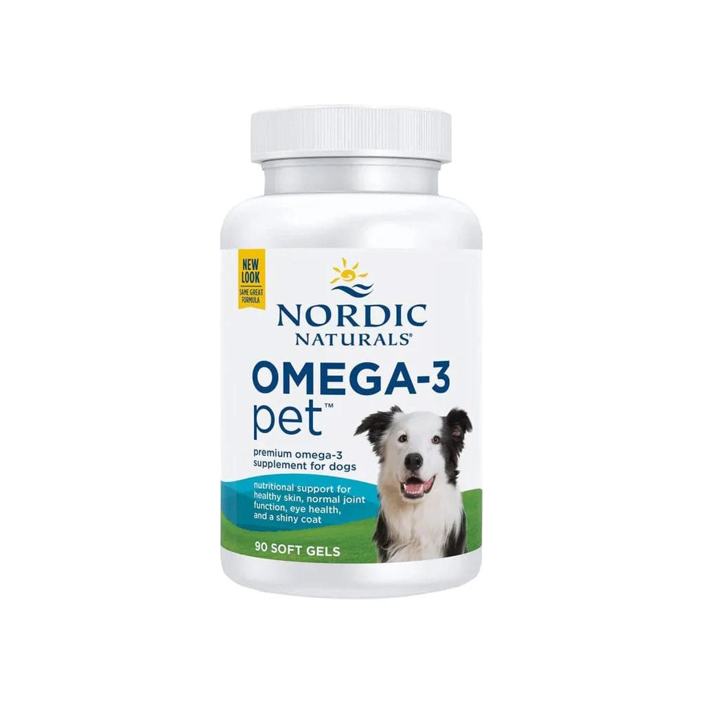
Third-party-tested omega-3 softgels (EPA and DHA) that support skin, coat, and joint health in dogs. Fish oil is one of the first things many vets suggest for a dull, dry, or flaky coat.
Webvet may earn a commission when you click through to Chewy, at no extra cost to you.
Tick Bites and Other Look-Alike Bumps
Not every bump on a dog is a growth. A tick bite lump on a dog is one of the most common false alarms: after a tick detaches or is removed, the bite site often forms a firm, pea-sized granuloma, a knot of inflammatory tissue that can persist for several weeks before flattening out. If you find a small, firm bump in an area where you recently pulled a tick, part the fur and look for a central scab or tiny crater. Mark the spot, measure it, and recheck weekly; a true tick-bite granuloma should shrink, not grow.
Other lump impostors worth knowing:
- An embedded tick itself. An engorged tick feels like a smooth gray-brown bump attached to the skin. Look for legs at the base before assuming skin growth.
- Insect bites and stings. These appear suddenly, are often warm and mildly swollen, and resolve within days. Hives can produce dozens of soft bumps at once.
- Vaccine-site lumps. A firm, small lump at an injection site within days of a vaccine is a common inflammatory reaction. Most resolve within a few weeks; per widely used veterinary guidance, any injection-site lump that is still growing after a month, exceeds 2 centimeters, or persists past three months should be biopsied.
- Scar tissue and calluses. Elbows and other pressure points build thickened, hairless pads that owners sometimes mistake for flat tumors.
If the bump traces back to a recent tick and you want to compare what bites look like as they heal, the tick-bite granuloma pattern is distinct from the growing, changing behavior of a true mass. When in doubt, apply the one-month rule: anything still present and unchanged or growing at the 30-day mark goes to the vet.
Emergency Signs: When a Lump Means Same-Day Vet Care
Most of these emergency presentations turn out to be abscesses or hematomas rather than cancer, and both are very treatable, but both are also painful and get worse without drainage or repair. The genuinely malignant emergencies, such as a ruptured splenic mass presenting as sudden belly distension and weakness, are time-critical in hours. The cost of an unnecessary same-day visit is a modest exam fee. The cost of waiting on a true emergency can be your dog's life. When your gut says something changed fast, trust it.
What the Vet Will Do: FNA vs Biopsy and Typical Costs
Knowing what happens at the appointment removes most of the dread. For the majority of skin lumps, the workup follows a standard escalation.
- Physical exam and history. Your vet measures the lump, checks its mobility and texture, palpates the regional lymph nodes, and asks how fast it appeared. Bring your photo log; growth history genuinely changes decisions.
- Fine needle aspiration (FNA). The first-line test for most lumps. The vet inserts a thin needle, no larger than a vaccine needle, draws cells, and examines them on a slide, often in-house within minutes, or sends them to a clinical pathologist. FNA needs no sedation for most dogs, and it reliably identifies lipomas, mast cell tumors, abscesses, and many other common answers. Typical cost runs roughly $75-$250 including in-house cytology, and more like $150-$400 when slides go to an outside pathologist.
- Biopsy. When aspiration is inconclusive, or when the surgical plan depends on the exact tumor type and grade, the vet takes a tissue sample under sedation or anesthesia, either a punch or incisional biopsy of part of the mass or an excisional biopsy removing the whole lump. Histopathology on the tissue gives the definitive diagnosis and grade. Typical cost, including sedation and lab fees, runs roughly $300-$800, more for masses in difficult locations.
- Staging tests when needed. If cytology or biopsy finds a malignancy, bloodwork, lymph node sampling, and imaging such as chest X-rays or abdominal ultrasound check whether the tumor has spread. Board-certified oncologists affiliated with the American College of Veterinary Internal Medicine describe these staging steps in their owner-facing resources (ACVIM veterinary specialty information).
The practical takeaway: the first diagnostic step is fast, low-stress, and modestly priced. Owners often delay the vet visit fearing a huge surgical estimate, when the actual first appointment is usually an exam plus a needle test. And if the lump is malignant, that early aspirate is what makes the eventual surgery smaller, cheaper, and more likely curative. For context on outcomes when testing does find something serious, see our guides to mast cell tumor life expectancy and dog lymphoma life expectancy: both make the same point from different angles, that stage at diagnosis drives prognosis.
How to Monitor a Lump at Home: The Measure-and-Photo Log
For lower-risk lumps your vet has examined, or new lumps awaiting an appointment, vague watching is not monitoring. Use a measure-and-photo log so change is measured, not guessed. Human memory reliably underestimates slow growth; a log does not.
- Measure the lump with a ruler or soft tape. Record length and width in millimeters. Calipers are ideal but a ruler is fine.
- Photograph it beside a coin. Part the fur, place a coin next to the lump for scale, and shoot from the same angle each time in good light.
- Note the feel. Soft or firm, movable or fixed, and whether your dog reacts to gentle pressure.
- Log the date and location on a simple body map. A rough dog outline with an X per lump prevents the multi-lump confusion common in senior dogs.
- Repeat every 30 days. Do a monthly check of the whole dog while you are at it, running flat hands over the body, legs, tail, and under the jaw.
- Escalate on change. Any growth beyond a couple of millimeters, any new redness, ulceration, or pain, or any change in feel moves the lump back up the triage table and into the vet's office.
Bring the log to every appointment. A lump documented at 12 mm in March and 19 mm in June makes the aspirate-versus-monitor decision for you, and it is exactly the evidence your vet wishes every owner brought.
Never Squeeze, Pop, or Remove a Lump Yourself
This warning is load-bearing enough to earn its own section. Do not squeeze a lump to see what comes out. Do not lance a suspected cyst or abscess with a needle. Do not tie off a skin tag with thread or floss, and do not use any online remedy that promises to draw out or dissolve a growth.
Each of these home interventions has a specific harm attached:
- Squeezing a mast cell tumor can trigger mass degranulation, a histamine flood that causes dramatic local swelling and, in severe cases, body-wide effects on blood pressure and the stomach lining.
- Squeezing a sebaceous cyst ruptures it under the skin, spilling irritant material that causes painful inflammation and infection, and usually guarantees the cyst returns.
- Lancing an abscess at home introduces new bacteria, misses the drainage and flushing the pocket actually needs, and skips the antibiotics and pain control that make treatment humane.
- Tying off skin tags causes slow, painful tissue death and open wounds, and if the growth was not actually a benign tag, it destroys the tissue a pathologist needed to see.
If a lump bursts or drains on its own, cover it loosely with a clean cloth, prevent licking, and get same-day veterinary care. The safe home intervention list for lumps has exactly three items: observe, measure, and photograph. A soft recovery cone or a snug recovery suit blocks licking far better than improvised bandaging while you wait for the appointment.
Lump on Dog FAQs
Our sister site Petful walks the same decision from the owner's side in Suspicious Lump on Your Dog? When to Tell the Vet, a useful second read before your appointment.
Frequently Asked Questions
My dog has a golf-ball-size lump on the neck that appeared overnight. Is it an emergency?
Overnight swelling warrants a same-day vet call. True tumors almost never appear in hours, so a sudden golf-ball lump on the neck is usually an abscess from a bite or puncture, a hematoma, an insect sting reaction, or a suddenly reactive lymph node or salivary gland problem. Several of these are painful and get worse without drainage or treatment, and neck swellings carry the extra risk of interfering with breathing or swallowing. Call your vet the same day, and treat it as urgent if your dog is lethargic, feverish, off food, or having any trouble breathing.
Can I pop or squeeze a lump on my dog?
No. Never squeeze a lump on a dog, even one that looks like a pimple or cyst. If the lump is a mast cell tumor, squeezing carries a real degranulation risk, releasing histamine that causes rapid swelling and can affect the whole body. If it is a cyst, squeezing spills irritant material under the skin and causes painful infection, and if it is an abscess, squeezing pushes bacteria deeper. Popping also destroys cells a veterinarian needs for an accurate aspirate. Observe, measure, and photograph at home; leave needles and squeezing to the clinic.
How long should I wait before having a new lump checked?
Use the one-month, one-centimeter guideline many oncology services teach: any lump that is still present after a month, is larger than about a pea, or is growing should be tested with fine needle aspiration rather than watched. Move faster, within days, if the lump is red, ulcerated, painful, fixed in place, or growing quickly, and treat bleeding, overnight swelling, or interference with breathing or eating as same-day problems. There is no prize for waiting; early aspirates are quick, affordable, and turn scary unknowns into answers.

Editor
The Webvet Editorial Team is the in-house group of pet-care editors and writers behind Webvet, operated by Smart Pet Collective. The team researches, writes, and maintains Webvet's pet health, behavior, and medication content. Every article follows a defined editorial process: research from reputable veterinary and scientific sources, careful drafting, mandatory review of medical content by a credentialed veterinarian, and dated publication. Health and medication articles are medically reviewed by a licensed veterinary professional before they go live and are kept current over time.
Veterinarian · BVMS MRCVS
Dr. Pippa Elliott, BVMS, MRCVS, is a veterinarian with nearly 30 years of experience in companion animal practice. Dr. Elliott earned her Bachelor of Veterinary Medicine and Surgery from the University of Glasgow. She was also designated a Member of the Royal College of Veterinary Surgeons. Married with 2 grown-up kids, Dr. Elliott has a naughty Puggle named Poggle, 3 cats and a bearded dragon.


