Histiocytoma Dog Lump: Pictures by Stage and Why It May Disappear on Its Own
A histiocytoma is the benign button tumor of young dogs. See pictures by stage with a week-marked regression sequence, the age rule that separates watch-and-wait from biopsy, and when removal is needed.

BVMS MRCVS
Medically reviewed by Dr. Pippa Elliott, BVMS MRCVS · Last reviewed

This article contains affiliate links. Webvet may earn a commission when you buy through them, at no extra cost to you.
If you have just found a small, raised, pink bump on your young dog, there is a good chance you are looking at the classic histiocytoma dog owners know as the "button tumor." It appears fast, looks alarming, and is one of the very few lumps in veterinary medicine that usually cures itself. This guide shows you histiocytoma pictures by stage, including a week-by-week regression sequence, explains how a histiocytoma differs from a mast cell tumor, and tells you exactly when treatment is actually needed.
Here is the tension every owner feels: a histiocytoma is benign and usually goes away on its own, yet it can look nearly identical to a mast cell tumor, which is cancer. That is why the plan below is always the same: get the lump checked and confirmed first, then relax and watch it regress.
- 1A histiocytoma is a benign "button tumor" that most often appears on dogs under about 3 years old.
- 2The age rule: under about three years, spontaneous regression is the expected course; the same-looking lump on an older dog earns a fine needle aspirate or biopsy.
- 3Most histiocytomas regress on their own within 2 to 3 months.
- 4A histiocytoma can mimic a mast cell tumor by eye; only FNA cytology can tell them apart.
- 5See your vet for confirmation before watch-and-wait, and never squeeze or home-remove any lump.

Sponsored Lemonade pet insurance link. Webvet may earn a commission.
What Is a Histiocytoma in Dogs?
A histiocytoma is a benign skin tumor made of Langerhans cells, a type of immune cell that lives in the skin and patrols for invaders. When these cells over-multiply in one spot, they form a firm, raised, hairless, often bright pink dome. Veterinarians nickname it the button tumor because it really does look like a small button sitting on the skin, typically 0.5 to 2.5 centimeters across. The formal name is cutaneous histiocytoma, but "the button tumor of young dogs" is how most vets describe it to owners.
The signature features that make a vet think "histiocytoma" on sight are:
- A young dog. Most cases occur in dogs under about 3 years old, and it is one of the most common skin tumors of young dogs.
- Rapid appearance. The lump often seems to show up over one to four weeks, then stops growing.
- A solitary, hairless, pink to red dome. Usually a single lump, smooth at first, most often on the head, ear flaps, muzzle, neck, or limbs.
- Not painful. Most dogs ignore the lump completely unless it ulcerates or gets licked raw.
Breeds like Boxers, Bulldogs, Labrador Retrievers, Staffordshire Bull Terriers, and Dachshunds are reported to be predisposed, but any breed or mix can grow one. VCA Animal Hospitals describes the classic presentation and its benign behavior in its histiocytoma overview (VCA Animal Hospitals).
The age rule is the single most useful thing to remember from this entire article. In a dog under about three years old, a confirmed histiocytoma is expected to regress on its own. A histiocytoma-looking lump on an older dog is a different situation entirely: it earns an FNA or biopsy, because mast cell tumors and other malignancies mimic it in that age group.

 Sponsored
Want a vet's take on your dog's skin or coat?
Talk to a licensed vet at Dutch over video, often the same day. They can recommend treatment and prescribe it when it's appropriate.
Use code Pets30
Plans start at $11/month · covers up to 5 pets
- Licensed vets in most states
- Rx for 150+ conditions
- 4.7 on Trustpilot
What Causes Histiocytomas in Dogs?
What causes histiocytoma in dogs? Nobody has identified a single trigger, and when owners search histiocytoma in dogs causes or histiocytoma dog cause, the honest answer is that no diet, product, or exposure has been implicated. Histiocytomas arise from Langerhans cells, and the leading understanding is that they represent a localized overgrowth of these immune cells rather than a response to injury, allergy, or infection. They are not contagious, they are not caused by anything you fed or applied, and there is no evidence an owner can prevent them.
A few points owners ask about most:
- Is it hereditary? No specific gene has been proven, but the strong breed predispositions suggest genetics load the dice.
- Is it from a bug bite or vaccine? Histiocytomas are not linked to either, although insect bite reactions can look similar for a few days.
- Why young dogs? The favored theory is that the young immune system's Langerhans cells are more prone to this transient proliferation, which also explains why the immune system later destroys the lump on its own.
The Merck Veterinary Manual groups histiocytomas with the histiocytic skin diseases and notes their benign, self-limiting course in young dogs (Merck Veterinary Manual).
Histiocytoma Pictures by Stage, Including Regression
Owners search for pictures of histiocytoma in dogs for one reason: to compare the lump on their own dog against the real thing. The photographs below show the stages in order, from the first small pink dome through the ulcerated phase. Match your dog's lump against each stage, but remember that a photo match is a starting hypothesis, not a diagnosis.
Location changes what these photos show more than most owners expect. Pictures of histiocytoma in dogs on the leg or paw usually capture a tight pink button, while images of histiocytoma on a dog's face, head, or neck often look flatter and redder against thinner skin. Early stage pictures of a histiocytoma on the leg can pass for a bug bite in one photo and an obvious dome in the next, and pics of an ear-flap lump often show crusting first. Whatever picture of a histiocytoma in dogs you match against, the location does not change the biology: the same young-dog button tumor behaves the same way on a paw, a leg, or a muzzle.

The first photo above shows the early stage histiocytoma most owners find; the ulcerated histiocytoma photo below it shows the stage that follows in many dogs.


A collar-mounted sensor and app for activity, rest, sleep, respiratory rate, heart-rate trends, itch behaviors, and more.
- Builds a personalized baseline over time
- Tracks trends between veterinary visits
- Generates a month-long shareable report
Health monitoring for dogs and cats
Webvet may earn a commission when you click through to Maven Pet, at no extra cost to you.
Early Stage
An early stage histiocytoma is a small, smooth pink dome, usually well under 2.5 centimeters, with a hairless, shiny surface. It sits proud of the skin like a button and feels firm but freely movable. At this stage it is easy to mistake for a wart, an insect bite, or a small mast cell tumor, which is exactly why vets aspirate first. Common locations are the ear flap, muzzle, head, and front legs, including the paw.
Peak and Ulcerated Stage
Many histiocytomas ulcerate at their peak. The surface breaks open, weeps, crusts, and forms a scabbed surface that can look raw and angry. Counterintuitively, an ulcerated histiocytoma is often a lump that is beginning to die back: the immune attack that destroys the tumor also damages its surface. Ulceration is the stage where secondary infection and licking become real risks, so this is a see-your-vet moment even if the lump was already diagnosed.
Regressing Stage
In regression, the dome flattens, shrinks, and darkens or scabs before the skin gradually returns to normal. The full week-by-week photo sequence of a single regressing lump is in the next section, because watching regression happen is the part no text description does justice.
 Sponsored
Vet care that's always there
Unlimited video calls and messaging with licensed vets for less than a typical vet visit. One membership covers up to five pets.
Use code Pets30
Plans start at $11/month · covers up to 5 pets
- Licensed vets in most states
- Rx for 150+ conditions
- 4.7 on Trustpilot
Will It Go Away on Its Own? The Regression Timeline
Yes, in most young dogs a confirmed histiocytoma goes away on its own. Most histiocytomas regress spontaneously within 2 to 3 months, as the dog's own immune system, led by infiltrating lymphocytes, destroys the tumor (VCA Animal Hospitals). Some resolve in as little as four weeks; a minority take longer, and a lump that is not clearly shrinking by about the 3 month mark should go back to the vet.
Because owners almost never get to see regression actually happening, here is the same lump photographed early and late in the course:

Read the pair left to right: early in the course the lump is at full size, a taut pink dome. Late in the course the surface has scabbed, the dome has flattened, and what remains is a faint pink patch on its way to disappearing. Between those two points, expect the same stage-by-stage regression on your own dog: scabbing first, then flattening week by week, then fading. That is the pattern you are hoping to see, and photographing your dog's lump weekly is the easiest way to confirm it is following the script.
| Phase | Typical timing | What owners usually see |
|---|---|---|
| Growth | Weeks 1 to 4 | Lump appears and enlarges quickly, smooth pink dome |
| Plateau | Roughly weeks 3 to 6 | Growth stops; surface may ulcerate, weep, or scab |
| Active regression | Roughly weeks 5 to 10 | Lump softens, flattens, and shrinks; scabbing is common |
| Resolution | By about 2 to 3 months | Lump gone; faint pink mark or small scar fades over time |
Timing varies dog to dog; the point of the table is the direction of travel. A lump following this arc is behaving like a histiocytoma. A lump that keeps growing past a month, or that has not shrunk by three months, has broken the pattern and needs to be re-examined and usually removed.
Owners also ask how long a dog can live with a histiocytoma, and this is the happiest answer in veterinary oncology: a confirmed histiocytoma has no effect on lifespan at all. It is benign, it does not spread to organs or lymph nodes in its ordinary form, and once it regresses or is removed, it is gone. Recurrence at the same spot is rare, though a dog can occasionally grow a new one elsewhere. Your dog's life expectancy is exactly what it was before the lump appeared.
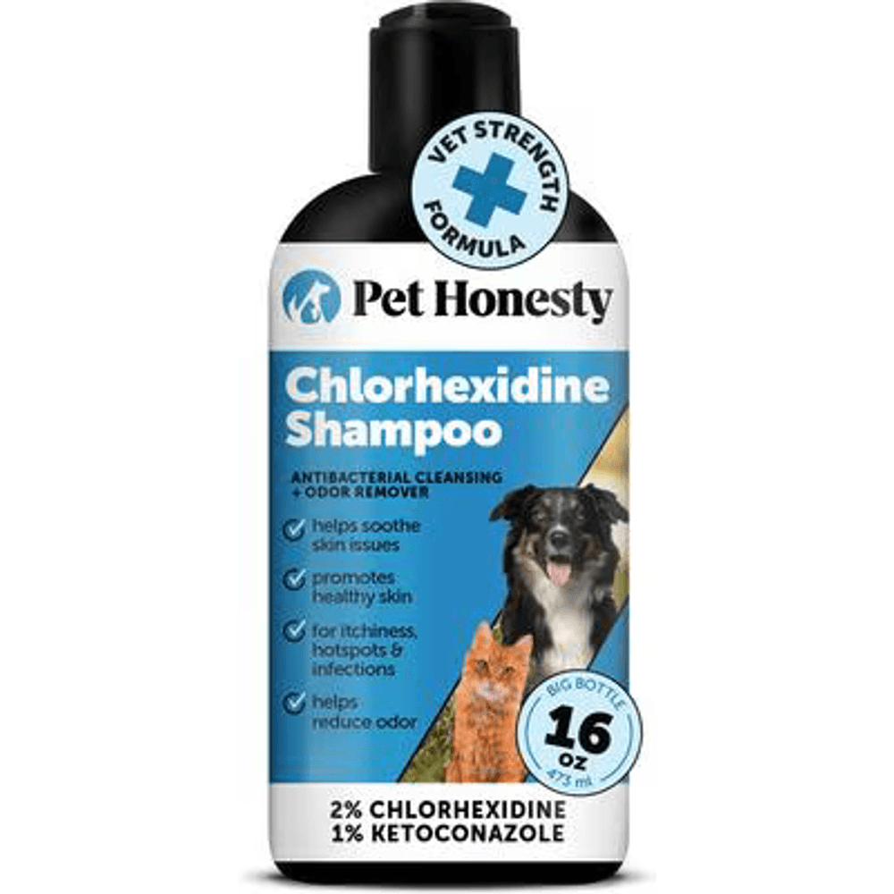
A vet-strength medicated shampoo with 2% chlorhexidine and 1% ketoconazole, the antifungal and antibacterial combination vets use to help clear ringworm and skin infections and to cut fungal spore shedding during treatment.
Webvet may earn a commission when you click through to Chewy, at no extra cost to you.
How Vets Confirm a Histiocytoma: FNA and Cytology
Fine needle aspiration is the quick, nearly painless test that settles the question. Your vet inserts a small needle into the lump, draws out a sample of cells, smears them on a slide, and examines them under the microscope. That microscopic exam is called cytology, and most dogs tolerate the whole process without sedation, often without even flinching.
On cytology, a histiocytoma shows sheets of uniform round histiocytes, sometimes with the lymphocytes that signal regression has already begun. A mast cell tumor looks completely different under the microscope, with granule-filled mast cells that stain distinctively. This is why FNA is so valuable: two lumps that are identical to the naked eye are unmistakably different on a slide.
Cytology occasionally comes back ambiguous, especially if the sample is thin or the lump is already regressing. In those cases your vet may repeat the aspirate, recommend removal with biopsy and histology, or have a pathologist review the slides. A histology report from an excised lump is the gold standard and doubles as the cure.
Histiocytoma vs. Mast Cell Tumor
This is the comparison that matters most, because it is the benign look-alike versus the most common malignant skin tumor of dogs. Both can be raised, pink, hairless, and fast-appearing. Mast cell tumors in dogs are famous shape-shifters, and a subset of them present exactly like a button tumor on a young adult dog.

Study the side by side pair above and you will see the problem: the two lumps overlap so much in appearance that even experienced veterinarians will not call it by eye. For a broader visual library of how mast cell tumors present, see our full set of mast cell tumor pictures.
| Feature | Histiocytoma | Mast cell tumor |
|---|---|---|
| Typical age | Usually under about 3 years | Usually middle-aged to older, average 8 to 9 years |
| Behavior | Benign, regresses within 2 to 3 months | Malignant, can spread; grade determines behavior |
| Look and feel | Firm, smooth pink dome; may ulcerate | Highly variable; can swell, shrink, redden day to day |
| Course over weeks | Stops growing, then shrinks | Persists, grows, or fluctuates; does not resolve |
| Fine needle aspiration | Uniform round histiocytes | Granule-filled mast cells |
| What happens next | Watch it regress, or remove if problematic | Surgical removal and staging, sometimes further treatment |
The single practical takeaway: appearance cannot rule cancer in or out, and the FNA settles it in minutes for a modest fee. No responsible watch-and-wait plan starts before that needle.
If the FNA Says Mast Cell Tumor
Sometimes the button-shaped lump on a young dog comes back from cytology as a mast cell tumor, and it is worth knowing that this is not a catastrophe. Mast cell tumors in young dogs are frequently low grade, and complete surgical removal is curative for the majority of low-grade tumors. Your vet will talk you through grading, margins, and whether any staging tests are sensible before surgery. The full picture, including grades, prognosis, spread, and treatment options, lives in our dedicated guide to mast cell tumors in dogs, which is the right next read the moment cytology changes the diagnosis.

A lightweight, padded fabric cone that gently blocks a pet from pawing, scratching, or rubbing a healing eye, wound, or hot spot, and it is far softer and less stressful than a hard plastic cone.
Webvet may earn a commission when you click through to Chewy, at no extra cost to you.
Histiocytoma in Older Dogs: When a Lump Deserves a Biopsy
Can a histiocytoma be cancerous in dogs? A true cutaneous histiocytoma is benign by definition, and that does not change with age. The danger is different: in an older dog, the odds that a histiocytoma-looking lump actually IS a histiocytoma drop sharply, while the odds it is a mast cell tumor, a histiocytic sarcoma, or another malignancy rise. The lump is not lying about what it is; the eye is simply unable to tell.
That is the second half of the plain age-based decision rule: young dog = likely regression, older dog = biopsy-worthy. In a dog under about three, a button-shaped lump is statistically likely to be the real benign article, and FNA confirmation plus watchful waiting is reasonable. In a dog past middle age, the identical lump earns an FNA at minimum, and often an excisional biopsy with histology, because the malignant mimics in that age group are treatable exactly when they are caught early.
One name that scares owners in search results is "malignant fibrous histiocytoma," an older label for a soft tissue sarcoma that is a completely different disease despite the similar name. Likewise, histiocytic sarcoma is a serious cancer of a related cell line, seen mostly in Bernese Mountain Dogs, Rottweilers, and retrievers, and it has nothing to do with the benign button tumor beyond ancestry. If a pathology report uses one of these terms, ask your vet to walk you through it rather than searching the scary phrase alone.
When Treatment or Removal Is Needed
How do you treat a histiocytoma in a dog? In most cases you do not: most need no treatment at all, because the expected course for a confirmed histiocytoma in a young dog is spontaneous regression within 2 to 3 months. The standard plan is FNA confirmation, then monitoring with photos every week or two while the immune system does the work. Treatment or surgical removal is actually needed only when the lump is ulcerated, infected, bothering the dog, or not regressing by about 3 months.
One search worth decoding here: owners looking for pictures of histiocytoma in dogs leg treatment are usually watching an ulcerated lump on a front leg or paw, and the photos in the stages section above show exactly what that looks like before and during care. A recovery suit with front-leg coverage or a loose sock protects it from licking while regression runs its course.
Surgical removal is the treatment when it is needed, and vets recommend it in a few specific situations:
- Ulcerated and not healing, bleeding repeatedly, or continuously oozing.
- Infected, with spreading redness, pain, or discharge that antibiotics and protection cannot control.
- Bothering the dog: constant licking, scratching, or a location like the paw or eyelid margin where irritation never stops.
- Not regressing by about 3 months, or still growing after the first month.
- Diagnosis in doubt, because excision plus histology answers the question definitively.
Removal is typically a short, routine surgery, and it is curative. In the meantime, a soft cone and keeping the surface clean protect an intact or ulcerated lump from the dog's own tongue, which is the most common cause of complications.
A word on home treatment, because the searches are full of it: apple cider vinegar does not treat histiocytomas, and neither do homeopathic remedies, salves, or "drawing" pastes. There is no evidence any topical home remedy shrinks a histiocytoma, and acidic or caustic products on an ulcerated lump cause chemical burns on top of the original problem. The honest version of home care is protection and patience: block licking, keep it clean and dry, photograph it weekly, and let regression happen on schedule.
Other Lumps It Could Be
Histiocytomas share the stage with a crowd of other canine lumps, and a wrong guess in either direction has a cost. Soft, slow-growing lumps in middle-aged dogs are often lipomas; wart-like clusters may be viral papillomas; firm skin masses can be sebaceous adenomas, cysts, or the malignancies covered above. Raw, red, moist skin lesions that appear overnight are frequently hot spots on dogs rather than tumors at all, though an ulcerated histiocytoma can imitate one. For a full triage walkthrough of the eight common lump types, what each feels like, and which ones need a same-week vet visit, start with our pillar guide to finding a lump on your dog.
Histiocytoma FAQs
Frequently Asked Questions
Do histiocytomas scab over?
Yes, histiocytomas commonly scab over, and scabbing is normal during regression and is often the first visible sign the lump is dying back. The surface may ulcerate, weep, crust, and scab repeatedly as the immune system destroys the tumor from within. The boundary to watch: a scabbed lump that is also shrinking is on course, while a lump that is infected or bleeding, smells bad, grows despite scabbing, or is being licked raw needs a vet visit rather than more waiting.
Is a histiocytoma dangerous for my dog?
No. A histiocytoma is benign once confirmed by your veterinarian, does not spread to other organs, and has no effect on your dog's lifespan. The only real danger lies in the confirmation step: because mast cell tumors can look identical, a lump that never got FNA confirmation carries the risk of being something more serious in disguise. Get the aspirate done, and after that the lump is a waiting game, not a threat.
How fast does a histiocytoma grow?
Fast, and that speed frightens owners more than anything else about it. A histiocytoma typically appears and reaches full size within one to four weeks, then stops growing entirely. That plateau is characteristic: benign button tumors surge and stall, while many malignant lumps keep enlarging steadily. A lump that is still actively growing after the first month does not fit the classic pattern and should be rechecked by your vet even if an earlier exam was reassuring.
Can a dog get more than one histiocytoma?
Usually a histiocytoma is solitary, and one lump in a young dog is by far the most common presentation. Occasionally a dog develops more than one at once, or grows a new one months to years after the first regresses, and Shar-Peis are noted for multiple lesions. Multiple lumps deserve extra caution because they can also signal other histiocytic diseases, so each new lump gets its own FNA rather than a shrug.

Editor
The Webvet Editorial Team is the in-house group of pet-care editors and writers behind Webvet, operated by Smart Pet Collective. The team researches, writes, and maintains Webvet's pet health, behavior, and medication content. Every article follows a defined editorial process: research from reputable veterinary and scientific sources, careful drafting, mandatory review of medical content by a credentialed veterinarian, and dated publication. Health and medication articles are medically reviewed by a licensed veterinary professional before they go live and are kept current over time.
Veterinarian · BVMS MRCVS
Dr. Pippa Elliott, BVMS, MRCVS, is a veterinarian with nearly 30 years of experience in companion animal practice. Dr. Elliott earned her Bachelor of Veterinary Medicine and Surgery from the University of Glasgow. She was also designated a Member of the Royal College of Veterinary Surgeons. Married with 2 grown-up kids, Dr. Elliott has a naughty Puggle named Poggle, 3 cats and a bearded dragon.


