Mast Cell Tumor Pictures in Dogs: What They Really Look Like
A labeled photo library of mast cell tumor pictures in dogs: all six presentations, look-alike comparisons with histiocytomas, lipomas, and warts, plus the appearances that mean a same-day vet visit.

BVMS MRCVS
Medically reviewed by Dr. Pippa Elliott, BVMS MRCVS · Last reviewed

This article contains affiliate links. Webvet may earn a commission when you buy through them, at no extra cost to you.
These mast cell tumor pictures in dogs show every common presentation of the most frequently diagnosed skin cancer in dogs, photographed and labeled so you can compare each one against the lump you just found. Mast cell tumors are notorious shape-shifters. One looks like a small pink pimple, another like an angry ulcerated sore, and a third hides under the skin feeling exactly like a harmless fatty lump. That is why veterinarians call the mast cell tumor "the great pretender," and it is why a photo library organized by presentation is more useful than any single textbook image.
This gallery is built the same way as our dog eye ulcer photo library: every claim is tied to a labeled photograph, look-alikes are shown side by side instead of described in the abstract, and the appearances that mean a same-day vet visit are flagged in their own section so you never have to guess whether tonight is soon enough.
One boundary before the first photo, because it is the most important sentence on this page: no picture, including the ones below, can diagnose a mast cell tumor. Mast cell tumors mimic benign lumps so convincingly that even veterinary oncologists will not call one from appearance alone. A photo match tells you what to do next, not what your dog has. The confirming test, fine needle aspiration, takes minutes and is covered at the end of this guide.
- 1Mast cell tumors are the most common skin cancer in dogs and can look like almost any lump: red and raised, ulcerated, or soft under the skin.
- 2Use the labeled photos below to match your dog's lump to a presentation, then match it against the four common look-alikes.
- 3A lump that swells, shrinks, or changes color overnight is behaving like a mast cell tumor and needs a vet promptly.
- 4No photo can diagnose or rule out cancer. A fine needle aspirate at your vet is the only way to know.

Sponsored Lemonade pet insurance link. Webvet may earn a commission.
What a Mast Cell Tumor Looks Like on a Dog: Photo Gallery
So what does mast cell cancer look like on a dog? Most often it looks like a raised, red or pink lump on or just under the skin, anywhere from a few millimeters to several centimeters across. It may be smooth or ulcerated, haired or hairless, firm or squishy. Roughly half appear on the trunk and belly, another quarter on the legs and paws, and the rest on the head, neck, and elsewhere. This page is a labeled real-photo gallery covering all six presentations: raised red, ulcerated, subcutaneous, early stage, mouth, and paw. The three in this first block, raised red, ulcerated, and early stage, account for the majority of cutaneous mast cell tumors owners find at home; the location-specific forms follow in the next gallery. Unlike the pharma treatment-journey galleries that dominate this search, every picture of a mast cell tumor on a dog here carries ID guidance and a clear cue for when the appearance means a same-day vet visit.
As you scroll, compare three things against your dog's lump: the surface (smooth skin, hairless shine, or an open ulcerated surface), the color (pink, red, or darker), and the border (a discrete bump versus a diffuse swelling). Note what matches and what does not; the pictures of mast cell tumor in dogs below are organized so you can check each feature in turn, and the same photos of these tumors double as a reference when the vet asks how the lump has changed. Your vet will ask exactly these questions, and photos of your dog's actual lump taken today give the appointment a head start.
A quick note on who gets these tumors, because it frames every photo that follows. Mast cell tumors account for roughly one in five canine skin tumors, making them the single most common skin cancer vets diagnose in dogs. Most affected dogs are middle-aged, around 8 to 9 years old on average, but they turn up in dogs of every age, including the young dogs discussed in the histiocytoma comparison later on. There is no reliable way to prevent them, so recognition is the whole game: the photos below are the difference between a tumor found at pea size and one found at plum size.
Raised Red Mast Cell Tumors
The classic presentation is a raised red bump sitting on top of the skin. It often starts hairless or loses hair as it grows, which gives it a smooth, shiny, inflamed look. Many owners first mistake it for a hive, an irritated insect sting, or a hot spot that will not resolve.

The raised red form is usually a discrete dome you can trace with a fingertip. Unlike a hive, it does not fade in a few hours, and unlike a hot spot, the dog usually has not been chewing the area raw. The redness comes from the histamine the tumor's mast cells leak into surrounding tissue, which also explains why these lumps are often itchy and why some dogs suddenly start licking one spot.

Look closely at the skin around the lump in the photo above. That surrounding flush is a useful tell. When a mast cell tumor is handled, rubbed, or scratched, the skin around it can redden and swell within minutes. Veterinarians call this Darier's sign, and it is strongly suggestive of a mast cell tumor rather than a benign growth. If stroking the lump makes it and the skin around it visibly angrier, tell your vet exactly that.

Breed matters when you are weighing how seriously to take a red bump. These tumors are heavily overrepresented in:
- Boxers, Boston Terriers, French Bulldogs, English Bulldogs, and Pugs
- Labrador Retrievers and Golden Retrievers
- Rhodesian Ridgebacks and Weimaraners
- Shar-Peis, which tend to develop more aggressive forms at younger ages
If your dog is on that list, any new red bump deserves an aspirate rather than a wait-and-see approach, even if it looks small and harmless in the mirror-image of the photos above.
Ulcerated Mast Cell Tumors
Some mast cell tumors outgrow their blood supply or are traumatized by licking, and the surface breaks down into an open sore. An ulcerated surface on a lump is never a normal finding, and it is one of the appearances covered again in the same-day section further down. Until the appointment, a soft recovery cone keeps licking from turning an irritated surface into an open one.

An ulcerated mast cell tumor typically looks like a raised mass with a wet, raw, or crusted crater in the middle. Owners often assume the dog "scratched a scab open," but the sequence is usually the reverse: the tumor ulcerated on its own, and the dog started licking because it became painful and itchy.

Bleeding is common with this presentation because degranulating mast cells release heparin, a natural blood thinner, directly into the tissue. That is why an ulcerated mast cell tumor can ooze far longer than a simple scrape of the same size, and why these lumps often re-bleed at night after the dog lies on them.

The larger, chronic ulcerated form in this photo shows what several weeks or months of growth can look like. The surrounding skin thickens and darkens, the crater widens, and secondary infection is common. Even at this stage, treatment can succeed, but every week of delay makes clean surgical removal harder. If your dog's lump resembles any of these three ulcerated photos, skip the watch-and-wait entirely.
Early Stage Mast Cell Tumors
The early stage presentation is the one this page most wants you to recognize, because small, low-grade mast cell tumors that are removed promptly are very often cured with surgery alone.

An early stage mast cell tumor is usually a small pink or flesh-colored bump, often no bigger than a pea. It can genuinely pass for a pimple, a wart, or an insect bite, which is exactly how so many get dismissed for months. The giveaway is persistence: a pimple or bite resolves within days, while an early mast cell tumor stays, fluctuates, or slowly grows.

Most early tumors are found by hand, not by eye. You feel a small firm bump while petting, part the coat, and find something like the photo above. This is why a monthly hands-on lump check, running your fingers slowly over the whole body, catches mast cell tumors at their most curable stage. A useful routine takes two minutes:
- Start at the muzzle and lips, then work over the head and ears
- Run flat palms down the neck, chest, and both sides of the trunk
- Check the belly, groin, and armpits, where coat hides small bumps best
- Finish with each leg, each paw, and between every pair of toes
Anything new gets photographed beside a coin and added to a lump map, a simple body outline where you mark each finding with its date and size. Vets love these maps because "new since last month" is more diagnostic than anything a single exam can show.

A collar-mounted sensor and app for activity, rest, sleep, respiratory rate, heart-rate trends, itch behaviors, and more.
- Builds a personalized baseline over time
- Tracks trends between veterinary visits
- Generates a month-long shareable report
Health monitoring for dogs and cats
Webvet may earn a commission when you click through to Maven Pet, at no extra cost to you.

Note that early lesions are not always domes. The photo above shows a flatter, slightly reddened patch, another legitimate early presentation. Any new skin change that persists beyond two weeks earns a vet visit under the widely used guideline oncologists teach: if a lump is bigger than a pea or has been there more than a month, aspirate it. Waiting costs nothing only when the lump is benign, and no photo can tell you that in advance.
How Big Is a Mast Cell Tumor? A Size Reference

Photos without scale mislead. The size reference above shows a typical early mast cell tumor beside a quarter: most owner-found tumors are between the size of a pencil eraser and a quarter at first discovery. Size alone does not predict grade, a small tumor can be high grade and a large one low grade, but size does affect surgery. Tumors under about 2 to 3 centimeters are far easier to remove with the wide margins mast cell tumors require. Measure your dog's lump against a coin today and write the date down; that measurement becomes the baseline for the rapid-growth warning sign covered below.

 Sponsored
Want a vet's take on your dog's skin or coat?
Talk to a licensed vet at Dutch over video, often the same day. They can recommend treatment and prescribe it when it's appropriate.
Use code Pets30
Plans start at $11/month · covers up to 5 pets
- Licensed vets in most states
- Rx for 150+ conditions
- 4.7 on Trustpilot
Gallery by Presentation and Location
Location changes both what a mast cell tumor looks like and how urgently it should be evaluated. The three location-specific presentations below, subcutaneous, mouth, and paw or leg, are the ones owners most often misread, and two of them carry higher stakes than the classic skin form.
Location also carries prognostic weight on its own. Oncology texts consistently flag tumors of the muzzle, mouth, nail bed, and groin as more likely to behave aggressively than trunk tumors of the same size, while subcutaneous tumors as a group tend to behave more gently than their skin-surface counterparts. None of that changes what you do at home, every location gets the same needle test, but it explains why the urgency advice below is not uniform across the three galleries.
Subcutaneous Mast Cell Tumors
A subcutaneous mast cell tumor grows in the fat layer under the skin rather than in the skin itself. There is often nothing to see at all, only something to feel: a soft, sometimes squishy lump that slides slightly under your fingers.

The danger of the subcutaneous form is what it imitates. A soft, movable lump under normal skin is exactly how a lipoma, the benign fatty tumor of older dogs, presents. Many subcutaneous mast cell tumors are dismissed for months as "just another fatty lump" precisely because the skin over them looks perfect, as in the photo above.

Palpation gives hints but never answers. Subcutaneous mast cell tumors are sometimes slightly firmer or more fixed than a classic lipoma, and they may fluctuate in size, which lipomas do not do. But studies of aspirated lumps show that vets themselves cannot reliably distinguish the two by feel, which is why the standard of care is to aspirate every new subcutaneous mass rather than assume lipoma.

The clipped view above shows the subtle skin dimpling a deeper subcutaneous tumor can cause. If you can see any surface change over a soft lump, mention it to your vet. The practical rule from this section is simple: a soft under-skin lump earns the same needle test as a red surface lump, because the reassuring feel of the subcutaneous form is exactly what makes it dangerous.
Mast Cell Tumors in a Dog's Mouth
A mast cell tumor in a dog's mouth or on the muzzle and lips is less common than the skin form, but it matters more, because oral and mucous-membrane mast cell tumors behave more aggressively and spread to lymph nodes earlier.

Mouth-area tumors usually appear along the lip margins, the muzzle, or the gums as a pink to red raised nodule, like the lip-margin example above. Owners often find them while brushing teeth or when the dog starts dropping food, pawing at the mouth, or drooling on one side.

Inside the mouth, as in the gum-line photo, a mast cell tumor can resemble a benign epulis, an abscessed tooth root swelling, or simple gum inflammation. Any distinct mass on the gums deserves prompt evaluation regardless of what it turns out to be, and mast cell tumor is one of the possibilities your vet will rank highly in this location.

Webvet may earn a commission when you click through to Chewy, at no extra cost to you.

Muzzle tumors like the one above sit in the zone veterinary oncologists treat with the most caution. Because tumors here reach the local lymph nodes early, a muzzle or oral mast cell tumor is a book-the-appointment-this-week finding even when it is small and quiet-looking. If your dog's lump matches this trio of photos, do not apply the two-week persistence rule from the early stage section; call sooner.
Mast Cell Tumors on the Paw and Leg

Paw and leg mast cell tumors are easy to misread as interdigital cysts, licked-raw allergy sores, or embedded foreign bodies like grass awns. The between-the-toes example above shows why: constant licking keeps the surface raw and inflamed, which looks exactly like the allergy sores many dogs get in the same spot. If your dog will not leave a paw lump alone, a recovery suit or a loose bootie protects it until the visit.

Lower-limb tumors like this hock example carry a practical surgical problem: there is very little spare skin on a dog's lower leg, so the wide margins mast cell tumor removal requires are hard to achieve. That is a second reason paw and leg lumps should be tested early, while they are small enough to remove cleanly.

A single swollen toe, as shown above, is a presentation owners almost never connect to cancer. It is usually blamed on a sprain or a torn nail. A toe that stays swollen beyond a week, without a known injury, deserves an exam, and your vet may recommend imaging along with the aspirate because toe tumors can involve bone. For the broader question of what else causes limb and paw lumps, our guide to other lumps and bumps on dogs walks through the full triage list with photos.
 Sponsored
Vet care that's always there
Unlimited video calls and messaging with licensed vets for less than a typical vet visit. One membership covers up to five pets.
Use code Pets30
Plans start at $11/month · covers up to 5 pets
- Licensed vets in most states
- Rx for 150+ conditions
- 4.7 on Trustpilot
Look-Alikes: Photos Compared Side by Side
What can be mistaken for a mast cell tumor in dogs? Four look-alikes cause most of the confusion:
- Histiocytoma, the benign "button tumor" of young dogs
- Lipoma, the soft fatty lump of middle-aged and older dogs
- Warts (papillomas) and other benign skin tags
- Bug bites, stings, and hives
Every one of these can be a dead ringer for a mast cell tumor, and a mast cell tumor can imitate every one of them. Few photo guides put these four common look-alikes side by side, so owners end up with five open tabs trying to compare. The look-alike photo pairs below fix that: each panel shows a confirmed mast cell tumor on the left and its look-alike, histiocytoma, lipoma, wart, or bug bite, on the right, photographed at similar scale.

The histiocytoma vs mast cell tumor comparison is the hardest one on this page, and the two photos above show why: both are pink-red, raised, hairless domes. The histiocytoma button is classically a fast-appearing dome on the ear, face, or leg of a dog under about 3 years old, and most regress on their own within a few months. But age is a probability, not a rule. Young dogs get mast cell tumors too, and the two are visually indistinguishable often enough that every "button tumor" should be confirmed by aspirate rather than assumed. Our full histiocytoma guide covers the regression timeline in detail.

The lipoma soft-lump comparison matters most for owners of older dogs. A lipoma is soft, oval, slow-growing, and slides under the fingers; the subcutaneous mast cell tumor in the left panel presents the same way. The one behavioral difference worth watching: lipomas essentially never fluctuate in size, while mast cell tumors swell and shrink. A "fatty lump" that changes week to week has broken the lipoma script. The safe path with any new soft mass is the one our lipoma guide recommends: aspirate once, then monitor with measurements.

The wart comparison is easier when the wart has its classic cauliflower texture, as in the right panel. True viral papillomas are usually rough-surfaced, pale, and often multiple, especially around the mouths of young dogs. The trap is the smooth wart and the smooth early mast cell tumor, which can be twins. Surface texture separates the textbook cases; the aspirate separates the rest. Our guides to dog warts and dog skin tags show the full range of benign surface growths for comparison.

The bug bite comparison comes down to time. Bites and hives, like the cluster in the right panel, appear suddenly, often in multiples, and resolve within hours to a couple of days. A mast cell tumor can look identical on day one, red, raised, itchy, but it is still there on day five. If you are watching a "bite" that has not resolved within a week, reclassify it as an unidentified lump and book the aspirate. Tick bites have their own distinctive look, compared photo by photo in our guide to what tick bites look like on dogs.
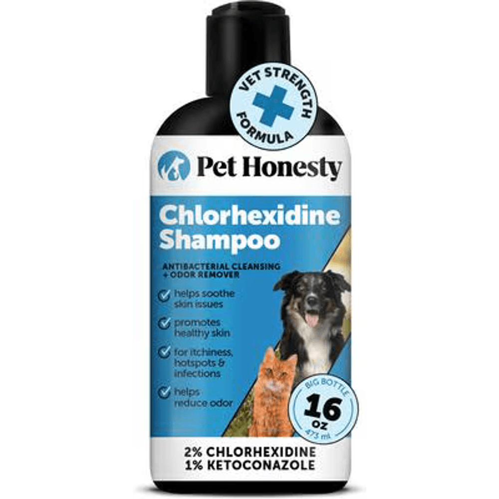
A vet-strength medicated shampoo with 2% chlorhexidine and 1% ketoconazole, the antifungal and antibacterial combination vets use to help clear ringworm and skin infections and to cut fungal spore shedding during treatment.
Webvet may earn a commission when you click through to Chewy, at no extra cost to you.
| Look-alike | Typical feel | Growth pattern | Color and surface | How it differs from an MCT | Urgency |
|---|---|---|---|---|---|
| Histiocytoma | Firm dome | Appears fast, then often regresses over weeks | Pink-red, hairless, smooth | Mostly dogs under 3; regression is typical | Aspirate to confirm; recheck monthly |
| Lipoma | Soft, movable | Very slow, steady | No surface change, normal skin | Never fluctuates in size day to day | Aspirate once, then measure and monitor |
| Wart (papilloma) | Firm, rough | Slow; may be multiple | Pale, cauliflower texture | Rough texture; often around mouth of young dogs | Vet check at next routine visit |
| Bug bite or hive | Soft swelling | Sudden onset | Red, may be multiple | Resolves within hours to days | Vet same day only if face swells or spreads |
| Mast cell tumor | Any of the above | Persists; may swell and shrink | Any; may ulcerate | Fluctuation, Darier's sign, persistence | Aspirate promptly; same-day if red-flag signs |
Is a mast cell tumor cancer in dogs? Yes. Every mast cell tumor is a true cancer, and the question is only its grade: many are low grade and curable with surgery alone, while a minority are high grade and spread. That is precisely why the look-alike table ends every row at the same place. Appearance sorts probabilities; only cytology sorts cancer from not-cancer. One other genuine cancer belongs on your radar in this section: cutaneous lymphoma can also mimic red patches and nodules, and our cutaneous lymphoma photo guide shows how its scaly, patchy early stage differs from the discrete lumps above.
The It Changed Overnight Pattern: Degranulation
If one behavior separates mast cell tumors from every look-alike on this page, it is this: they change size, fast, in both directions. Owners describe a lump that "doubled overnight," then a week later swear it is shrinking. Both reports are usually accurate, and the explanation is degranulation.
Mast cells are immune cells packed with granules of histamine and other inflammatory chemicals. When the cells in a tumor are disturbed, by rubbing, scratching, pressure from lying down, or nothing identifiable at all, they dump those granules into the surrounding tissue. The histamine release causes sudden swelling, redness, and itch. When the flare subsides, the lump deflates toward its previous size. That swell-and-shrink cycle is the degranulation swelling pattern, and no benign look-alike does it.

Degranulation is also the reason your vet will likely give an antihistamine such as diphenhydramine, and sometimes a stomach protectant, before removing a confirmed mast cell tumor. Blocking histamine at its receptors blunts the flare that surgical handling would otherwise trigger. Knowing that helps you read the photos in this section correctly: the swelling you see is a chemical event layered on top of the tumor, not the tumor itself growing.
The two-frame sequence above shows how the same tumor can look roughly twelve hours apart. In the first frame it is a modest pale bump; in the second it is red, tense, and nearly twice the diameter after an overnight flare. Seeing it on a single tumor matters, because this is the single most misread mast cell tumor behavior. The cruel part of the pattern is the shrink phase: owners watch the lump deflate and conclude it is healing, when a shrinking lump after a flare is actually evidence for a mast cell tumor, not against one.
The at-home takeaway is a log, not a guess. Photograph the lump next to a coin today, and again any morning it looks different. Three photos a week apart showing real size fluctuation are among the most useful things you can bring to an appointment.
Photos That Mean Same-Day Vet Care
Most lumps in this library justify a prompt appointment within days. The three photographed appearances in this section are different: they justify a call today, and an emergency visit tonight if your regular clinic cannot see you. This list is the reason this page exists, so treat it as load-bearing.
When you call, say the words that move you up the triage list: "my dog has a lump that is bleeding," or "a lump that visibly grew this week," or "several new lumps at once." Front-desk staff triage by phrase, and a vague "I found a bump" lands you in the routine queue that the appearances below should skip.

An ulcerated surface that is actively bleeding, like the photo above, is same-day territory for two reasons. First, the heparin mast cells release keeps these wounds oozing far longer than normal skin injuries, and dogs can lose meaningful blood from a large tumor overnight. Second, an open tumor surface infects easily. Cover it loosely with a clean non-stick dressing if the dog allows, prevent licking, and get seen today.

Visible rapid growth is the second same-day flag. The photo above shows a tumor against a coin with the previous week's outline marked; the difference is obvious without measurement. Growth you can see across days, as opposed to the slow drift of months, correlates with higher-grade behavior, and higher-grade tumors are the ones where treatment timing genuinely changes outcomes. A degranulation flare can mimic true growth, and same-day evaluation is how the two get told apart; that distinction is not one to make at home.

Multiple lumps appearing together, as in the photo above, is the third flag. Several simultaneous mast cell tumors can mean a dog is genetically primed to keep making them, or that an aggressive tumor has begun spreading through the skin and lymphatics. Either way, staging, checking lymph nodes and organs, moves up the priority list, and that starts with a same-day or next-morning exam rather than a wait for the next open slot.
- 1See a vet the same day for: an ulcerated surface that is bleeding, visible growth across days, multiple lumps at once, or a dramatic overnight degranulation flare.
- 2Add these whole-dog signs to the same-day list: vomiting, black tarry stool, pale gums, weakness, or collapse in a dog with a known or suspected mast cell tumor, because massive histamine release can ulcerate the stomach.
- 3For everything else that matches this page's photos, book the earliest routine appointment and ask for a fine needle aspirate. Days matter; hours usually do not.
Beyond the Photos: What the Vet Does Next
Here is why no photo can diagnose a mast cell tumor: everything on this page funnels to one test. Fine needle aspiration, FNA, is the confirming step that turns a suspicious photo match into an answer, and it is far smaller than most owners expect: a needle the size used for vaccines, no sedation in almost all cases, a few seconds per sample, and cytology results the same day if the clinic reads slides in-house or within a couple of days from a reference lab.

A few practical notes make the visit smoother. Ask for the aspirate at the same appointment as the exam rather than a recheck, since it rarely needs scheduling. Bring your photo log and coin measurements, because documented size fluctuation changes how aggressively a vet pursues an ambiguous cytology result. And if the first aspirate comes back inconclusive, which happens when a sample catches mostly blood or fluid, a repeat aspirate or a small biopsy is the next step, not a shrug.
Mast cell tumors are among the most cooperative cancers under the microscope. Their cells are round and stuffed with granules that stain vivid purple, so cytology confirms most cases without a surgical biopsy. What a photo can never show, and the aspirate begins to reveal, is the tumor's grade, the single biggest driver of everything that follows. From there, the roadmap runs through staging, surgical removal with wide margins, and sometimes radiation or medication, all of which belongs to its own guide. For grades, causes, treatment options, and what surgery involves, see our full guide to mast cell tumors in dogs: grades and treatment, which owns that half of the journey the way this page owns the photos.
What Happens After Diagnosis: Outlook
The honest answer to "what happens now" is that outcomes range from complete surgical cure, which is the most common result for low-grade tumors caught at the size shown in the early stage photos above, to genuinely serious disease for the minority of high-grade tumors. Where your dog lands depends on grade, location, completeness of removal, and how early you acted, which is exactly why this photo library leans so hard on early recognition. Survival numbers by grade and stage, and what they actually mean for an individual dog, deserve more than a paragraph, and they get it in our dedicated guide to mast cell tumor life expectancy by grade. Read it after the diagnosis, not instead of the appointment.
Mast Cell Tumor Picture FAQs
Frequently Asked Questions
Can a vet diagnose a mast cell tumor from a picture?
No. A photo alone cannot diagnose or rule out a mast cell tumor, and neither can a physical exam. Mast cell tumors imitate benign lumps too well, and benign lumps imitate them. A photo match on this page tells you the lump deserves testing, nothing more. The diagnosis comes from fine needle aspiration, where a needle sample of cells is examined under a microscope. It is quick, inexpensive, and usually needs no sedation, which is why vets recommend aspirating any new lump rather than judging it by eye.
How quickly can a mast cell tumor change in appearance?
Overnight. A mast cell tumor can swell overnight to double its size, turn red and angry, then deflate again over the following days as a degranulation flare subsides. That swell-and-shrink cycle is a hallmark of mast cell tumors, and shrinking does not mean benign: a lump that gets smaller after a flare has demonstrated mast cell behavior, not healing. Photograph the lump beside a coin whenever it changes and bring the photo series to your vet, because documented fluctuation is diagnostic gold.

Editor
The Webvet Editorial Team is the in-house group of pet-care editors and writers behind Webvet, operated by Smart Pet Collective. The team researches, writes, and maintains Webvet's pet health, behavior, and medication content. Every article follows a defined editorial process: research from reputable veterinary and scientific sources, careful drafting, mandatory review of medical content by a credentialed veterinarian, and dated publication. Health and medication articles are medically reviewed by a licensed veterinary professional before they go live and are kept current over time.
Veterinarian · BVMS MRCVS
Dr. Pippa Elliott, BVMS, MRCVS, is a veterinarian with nearly 30 years of experience in companion animal practice. Dr. Elliott earned her Bachelor of Veterinary Medicine and Surgery from the University of Glasgow. She was also designated a Member of the Royal College of Veterinary Surgeons. Married with 2 grown-up kids, Dr. Elliott has a naughty Puggle named Poggle, 3 cats and a bearded dragon.


