Mast Cell Tumor in Dogs Life Expectancy: By Grade and Stage
Honest, study-sourced answers on mast cell tumor in dogs life expectancy: survival by grade and stage, what the final stages look like, and a compassionate quality-of-life framework to walk through with your vet.

BVMS MRCVS
Medically reviewed by Dr. Pippa Elliott, BVMS MRCVS · Last reviewed

This article contains affiliate links. Webvet may earn a commission when you buy through them, at no extra cost to you.
If you are searching for mast cell tumor in dogs life expectancy, you are probably holding a biopsy report or waiting on one, and you need honest numbers instead of vague reassurance. Here is the truth up front: there is no single answer, because mast cell tumors range from lumps that are cured with one surgery to aggressive cancers measured in months. The two things that matter most are the tumor's grade and its stage, and this article walks through both with survival figures drawn only from published studies and veterinary institutions.
That range is wider than almost any other cancer in dogs. A dog with a low-grade tumor that is completely removed will usually go on to live a normal lifespan. A dog with a high-grade tumor that has already spread faces a much harder road. Knowing which situation your dog is in changes everything about the decisions ahead, which is why the sections below pair study-sourced survival tables by both grade and stage with a practical quality-of-life checklist, breaking life expectancy down by grade, by stage, and by whether treatment is pursued.

 Sponsored
Want a vet's take on your dog's skin or coat?
Talk to a licensed vet at Dutch over video, often the same day. They can recommend treatment and prescribe it when it's appropriate.
Use code Pets30
Plans start at $11/month · covers up to 5 pets
- Licensed vets in most states
- Rx for 150+ conditions
- 4.7 on Trustpilot
How long do dogs live with a mast cell tumor?
Most dogs with a low-grade mast cell tumor that is completely removed live a normal lifespan, with survival measured in years and many dogs effectively cured. Dogs with high-grade tumors have a median survival of less than four months without aggressive therapy, according to the grading study by Kiupel et al. 2011. Grade, stage, and mitotic index drive nearly every survival statistic in between.
- 1Low-grade mast cell tumors that are fully removed usually mean a normal lifespan, and many dogs are cured with surgery alone.
- 2High-grade disease is far more serious: Kiupel et al. 2011 reported a median survival under 4 months for high-grade tumors.
- 3Grade (how aggressive the cells look) and stage (how far the cancer has spread) are the two numbers that determine your dog's individual outlook.
- 4Life expectancy is a conversation to have with your veterinarian or a veterinary oncologist, using your dog's specific biopsy and staging results.

When your veterinarian talks about prognosis, they are combining several pieces of information from the pathology report:
- Grade: how abnormal and fast-dividing the tumor cells appear under the microscope
- Mitotic index: a count of actively dividing cells, one of the strongest single predictors of survival
- Stage: whether the tumor has spread to lymph nodes, the spleen, the liver, or the bone marrow
- Location: tumors on the skin of the body generally do better than tumors in the mouth, nail bed, or internal organs
- Completeness of removal: whether the surgeon achieved clean margins around the tumor
The mitotic index deserves special mention because the numbers attached to it are so stark. In a study of canine mast cell tumors, Romansik et al. 2007 reported that dogs whose tumors had a mitotic index of 5 or less had a median survival of about 70 months, while dogs whose tumors had a mitotic index above 5 had a median survival of roughly 2 months. That single line on a biopsy report separates dogs who live for years from dogs who live for weeks, which is why no one can quote a meaningful life expectancy until the pathology is back.

Sponsored Lemonade pet insurance link. Webvet may earn a commission.
So when someone asks how serious a mast cell tumor in dogs is, the honest answer is: it depends entirely on what the microscope shows. Roughly two thirds of canine mast cell tumors are low grade, per the Merck Veterinary Manual, which means the odds genuinely favor your dog until proven otherwise.
This guide is built for one job only. General MCT guides treat life expectancy as one short section and cite few or no published survival figures, so this page combines study-sourced survival tables by both grade and stage with a practical quality-of-life checklist. Below you get all three: a survival table by grade, a second table by stage, each figure tied to a published study or institution, and a quality-of-life checklist to walk through with your veterinarian.
The good news: low-grade tumors have an excellent outlook
Before we get into the harder numbers, this point deserves its own section because it gets buried on most pages: low-grade tumors carry an excellent prognosis, and the majority of dogs diagnosed with a mast cell tumor do well. Most cutaneous mast cell tumors are low grade, and complete surgical removal of a low-grade tumor is very often curative.

The published data backs this up consistently:
- In the original grading study by Patnaik et al. 1984, about 93 percent of dogs with grade I tumors were still alive roughly four years (1,500 days) after surgery.
- In the two-tier system study by Kiupel et al. 2011, more than 90 percent of low-grade tumors did not kill the dogs that had them; those dogs generally died of unrelated causes.
- Veterinary teaching institutions, including the Merck Veterinary Manual, describe complete excision of well-differentiated tumors as usually curative.
If your dog's biopsy comes back as a grade I or Kiupel low-grade tumor with clean surgical margins, the most likely outcome is that this lump never troubles your dog again. Your veterinarian will still recommend monitoring for new lumps, because dogs that develop one mast cell tumor are more likely to develop another, but a new tumor is a new event, not a spread of the old one. Regular lump checks at home, the same habit covered in our guide to finding a lump on your dog, become part of normal life rather than a source of dread.
Hold on to this while you read the rest of the article. The frightening numbers below describe the minority of cases, and they exist so that families facing high-grade or advanced disease get honest information too.
Understanding mast cell tumors: what drives the numbers
Mast cell tumors are cancers of mast cells, immune cells loaded with histamine and other inflammatory chemicals. That biology explains their strange behavior: tumors can swell and shrink day to day, and large or disturbed tumors can release histamine in bulk (degranulation), causing stomach ulcers, vomiting, and dangerous drops in blood pressure. It also explains why vets prescribe antihistamines and stomach protectants alongside cancer treatment.
This article stays focused on survival and end-of-life decisions. For everything else, including symptoms, causes, diagnosis by fine needle aspiration, surgery, radiation, chemotherapy, Palladia, Stelfonta, the cost of mast cell tumor removal in dogs, and questions about natural remedies for mast cell tumor in dogs (spoiler: none replaces surgery), our complete mast cell tumor guide covers the full journey from first lump to treatment planning. If you are still at the "what is this bump" stage, start there, because grade and stage cannot even be discussed until a tumor is sampled and diagnosed.

A collar-mounted sensor and app for activity, rest, sleep, respiratory rate, heart-rate trends, itch behaviors, and more.
- Builds a personalized baseline over time
- Tracks trends between veterinary visits
- Generates a month-long shareable report
Health monitoring for dogs and cats
Webvet may earn a commission when you click through to Maven Pet, at no extra cost to you.
What mast cell tumors look like
One reason mast cell tumors are called "the great pretenders" is that no appearance rules them out. They can look like a smooth pink bump, a wart, an insect bite, a bruise, or a soft fatty lump indistinguishable from a harmless lipoma. Young dogs often get benign histiocytomas that look nearly identical. Because your eye cannot grade a tumor, photographs are for recognition, not diagnosis. To see the full visual range, from tiny pink nodules to ulcerated masses, see our guide to what mast cell tumors look like, and let a needle sample make the actual call.
Life expectancy by tumor grade
Grade is assigned by a pathologist after a biopsy, and it is the single most quoted predictor of survival. The survival by grade table below draws every figure from a named published study or institution. Two systems are in use, and your report may cite either or both. The older Patnaik grading system (grades I, II, III) dates to 1984; the newer Kiupel grading system (low grade vs. high grade) was designed in 2011 to reduce the ambiguity of the middle grade.
| Grade | What it means | Published survival figures | Source |
|---|---|---|---|
| Grade I (Patnaik) | Well differentiated, least aggressive | About 93% of dogs alive at 1,500 days (roughly 4 years) after surgery | Patnaik et al. 1984 |
| Grade II (Patnaik) | Intermediate; behavior varies widely | About 47% of dogs alive at 1,500 days; many with complete excision are cured | Patnaik et al. 1984 |
| Grade III (Patnaik) | Poorly differentiated, most aggressive | About 6% of dogs alive at 1,500 days | Patnaik et al. 1984 |
| Low grade (Kiupel) | Favorable biology | More than 90% of dogs survive beyond 2 years; most die of unrelated causes | Kiupel et al. 2011 |
| High grade (Kiupel) | Aggressive biology | Median survival under 4 months | Kiupel et al. 2011 |
| Mitotic index 5 or less | Slow cell division | Median survival about 70 months | Romansik et al. 2007 |
| Mitotic index over 5 | Rapid cell division | Median survival about 2 months | Romansik et al. 2007 |
A few practical notes on reading that table:
- Grade 1 mast cell tumor dog life expectancy is, in most cases, simply the dog's normal life expectancy. With complete removal, recurrence and spread are uncommon.
- Grade 2 mast cell tumor dog life expectancy is the hardest to pin down because grade II is a mixed bag. This is exactly why the Kiupel system exists: a Patnaik grade II tumor that is Kiupel low grade usually behaves like a grade I, while a grade II that is Kiupel high grade behaves like a grade III. Ask your vet which Kiupel category your dog's grade II tumor falls into, along with its mitotic index.
- Grade III / high-grade tumors carry the hardest numbers in veterinary oncology, but they are not hopeless. With aggressive multimodal therapy (surgery, radiation, chemotherapy, or targeted drugs like toceranib), some dogs live well beyond the medians. In the pivotal Palladia trial, London et al. 2009 reported an objective response in about 43 percent of dogs with recurrent or metastatic mast cell tumors.
Medians are the middle of a curve, not a prediction for your individual dog. Half of dogs outlive every median in that table.
 Sponsored
Vet care that's always there
Unlimited video calls and messaging with licensed vets for less than a typical vet visit. One membership covers up to five pets.
Use code Pets30
Plans start at $11/month · covers up to 5 pets
- Licensed vets in most states
- Rx for 150+ conditions
- 4.7 on Trustpilot
Life expectancy by stage
Where grade describes how nasty the cells look, stage describes how far they have traveled. Staging tests typically include lymph node sampling, abdominal ultrasound of the spleen and liver, and sometimes bone marrow evaluation. The World Health Organization staging scheme for canine mast cell tumors runs from stage 0 to stage IV, and the study-sourced survival by stage table below summarizes what published data shows for each level, including stage 3.
| Stage | What it means | What published data shows | Source |
|---|---|---|---|
| Stage 0 | Incompletely removed tumor, no node involvement | Excellent with follow-up treatment of the surgical site | Merck Veterinary Manual |
| Stage I | One skin tumor, no lymph node spread | Best outlook; low-grade stage I disease is usually cured surgically | Patnaik et al. 1984; Merck Veterinary Manual |
| Stage II | One tumor with spread to the regional lymph node | Long-term survival still achievable when the node is treated; surgery plus radiation of the node has produced multi-year control in grade II disease | Chaffin and Thrall 2002 |
| Stage III | Multiple skin tumors, or a large deep tumor | Prognosis driven mostly by grade; multiple low-grade tumors can still do well | Merck Veterinary Manual |
| Stage IV | Distant spread: spleen, liver, blood, or bone marrow | Median survival around 110 days in dogs with systemic mast cell disease | Marconato et al. 2008 |
Two stage-specific questions come up constantly and deserve direct answers.

An orthopedic sofa bed with three bolstered sides for dogs who like to rest their head and joints against a cushion. The supportive foam base cradles hips and elbows, a comforting spot for an older or shaky dog to settle.
Webvet may earn a commission when you click through to Chewy, at no extra cost to you.
Stage 2 mast cell tumor in dogs prognosis
Lymph node involvement makes things more serious, but it is not the death sentence owners fear. When the affected node is removed or irradiated along with the primary tumor, studies such as Chaffin and Thrall 2002 have documented years of tumor control in dogs with grade II tumors and regional node spread. Many oncologists now treat the involved node aggressively for exactly this reason.
How long does a dog with mast cell tumors all over live?
It depends on what "all over" means, and the distinction matters enormously. Multiple separate skin tumors (stage III) are not the same as one cancer that has spread internally. A dog who grows several independent low-grade skin tumors over time can live for years, having each one removed as it appears; the Merck Veterinary Manual notes that multiple cutaneous tumors do not automatically worsen prognosis if each is low grade. True disseminated disease, where mast cells infiltrate the spleen, liver, or bone marrow, is stage IV, and there the published outlook is much shorter: Marconato et al. 2008 reported a median survival of about 110 days in dogs with systemic involvement. If your dog has many lumps, ask your vet to aspirate several and to stage the abdomen, because the answer changes the entire plan.
Stage 3 mast cell tumor in dogs prognosis
Stage 3 prognosis sits between those poles. The stage III label covers both multiple skin tumors and single large invasive ones, so oncologists lean on grade and mitotic index to predict behavior. A stage III dog with low-grade biology may still enjoy years; a stage III dog with high-grade biology tracks closer to the high-grade medians above.
Life expectancy without treatment
Some families cannot pursue surgery, whether because of cost, the dog's age, anesthetic risk, or other health problems. If that is you, you deserve honest information rather than judgment.
There is no large published study that followed untreated mast cell tumors to give a precise mast cell tumor dog life expectancy without treatment, so honest vets speak in patterns rather than exact figures. Those patterns come directly from tumor biology:
- Untreated low-grade tumors often grow slowly, sometimes remaining stable for months or longer. Some eventually ulcerate, itch, or interfere with movement depending on location. A watchful-waiting approach with regular measurement is a legitimate plan for a frail dog with a small low-grade tumor, decided with your vet.
- Untreated high-grade tumors typically progress quickly, over weeks to a few months, consistent with the sub-4-month median survival Kiupel et al. 2011 documented even among treated high-grade cases.
- Any untreated tumor can degranulate, releasing histamine that causes stomach ulcers, vomiting, loss of appetite, and in severe cases a dangerous blood pressure crash. This is the main way an untreated skin tumor makes a dog systemically ill.
Even when surgery is off the table, palliative care can meaningfully extend comfortable time. Prednisone can shrink mast cell tumors temporarily, and antihistamines (like diphenhydramine and famotidine-type stomach protectants) blunt the histamine effects. These are inexpensive oral medications, and your veterinarian can build a comfort-focused plan around them. Untreated never has to mean unsupported. Ask your vet about appetite stimulants such as Entyce (capromorelin) when meals start getting refused, and give an orthopedic bed the same priority as the pill bottle: comfortable rest is half of palliative care.
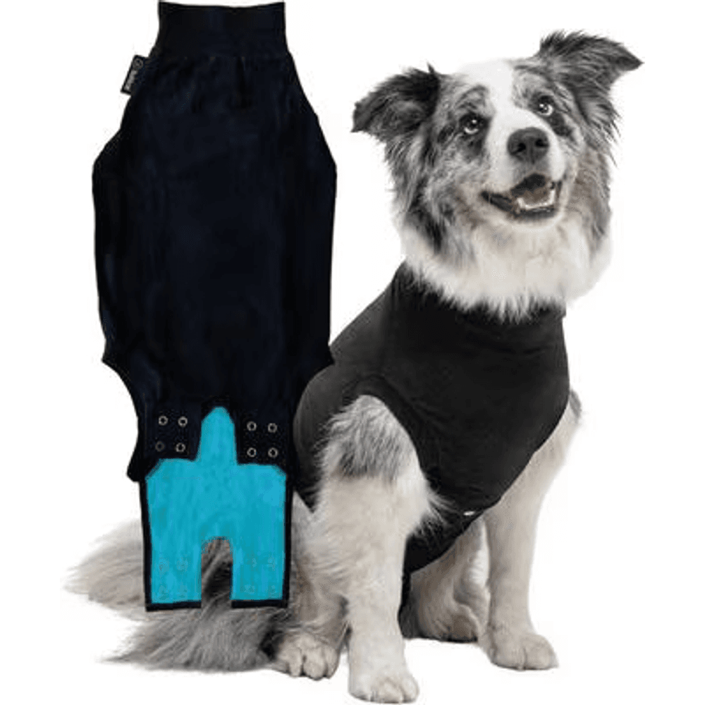
A breathable full-body recovery suit that covers a spay or abdominal incision, the vet-recommended cone alternative that lets a dog rest, walk, and go potty normally without licking or scratching the wound.
Webvet may earn a commission when you click through to Chewy, at no extra cost to you.
How fast do mast cell tumors spread?
Do mast cell tumors spread quickly in dogs? Speed tracks with grade, and the honest answer covers both ends. Low-grade tumors spread rarely and slowly: metastasis occurs in fewer than 10 percent of well-differentiated tumors, per figures compiled in the Merck Veterinary Manual, and many never spread at all. High-grade tumors are the opposite story, with the Merck Veterinary Manual citing metastatic rates for poorly differentiated tumors ranging from roughly 55 to 96 percent. When high-grade tumors spread, they can do so within weeks, moving first to the regional lymph node, then to the spleen and liver.
That spread pattern is worth knowing because it shapes monitoring:
- Regional lymph node is almost always the first stop, which is why your vet palpates and often aspirates the node nearest the tumor.
- Spleen and liver are the most common internal sites, screened with abdominal ultrasound.
- Bone marrow and blood involvement marks the most advanced disease.
- Lungs are usually spared, unlike most other cancers; chest metastasis is uncommon in mast cell disease.
Recurrence is the other clock owners watch. Mast cell tumor recurrence in dogs is mostly a story about surgical margins: tumors removed with clean margins seldom regrow at the site, while incompletely excised tumors may return, sometimes more aggressively. Recurrence at the surgical scar within months is a warning sign of aggressive biology, and any new lump on a dog with mast cell history should be aspirated promptly rather than watched.
A note on internal mast cell tumor in dogs: primary visceral mast cell disease (starting in the spleen, gut, or other organs rather than the skin) is thankfully uncommon but carries a substantially worse outlook than skin tumors, behaving like the stage IV disease described by Marconato et al. 2008. Vomiting, black stools, appetite loss, and lethargy in a dog with mast cell history warrant an ultrasound conversation with your vet.
What the final stages look like
If your dog's disease has advanced despite treatment, or you have chosen comfort care, knowing what to expect removes some of the fear. What are the final stages of mast cell tumors in dogs? In advanced disease, the tumor burden and circulating histamine begin to affect the whole body. Appetite is often the first thing to fade, degranulation episodes can cause sudden swelling or hives, and the final stages typically bring some combination of the changes below:
- Appetite loss and nausea, often from histamine-driven stomach ulcers; some dogs vomit, and vomit may contain digested blood that looks like coffee grounds
- Dark, tarry stools (digested blood from ulceration in the stomach or upper intestine)
- Visible tumor changes: rapid growth, ulceration, bleeding, or new lumps appearing faster than they can be addressed
- Swelling and redness around tumors, or hives and facial swelling after a degranulation episode
- Deep fatigue: sleeping most of the day, reluctance to walk, withdrawing from the family
- Weight and muscle loss even when eating, as advanced cancer consumes calories
- Pale gums, weakness, or collapse in the last days, from anemia, low blood pressure, or internal bleeding
Dogs in the final stage of mast cell disease usually decline through some combination of not eating, gastrointestinal bleeding, and exhaustion rather than sudden dramatic pain, though ulcerated tumors and stomach ulcers do hurt. This is how most dogs die from mast cell tumors when the disease itself is allowed to run its course, and it is the reason most veterinarians gently recommend euthanasia before the very end: the last stretch offers the dog little except discomfort.
If you are seeing several of these signs together, it is time for a frank conversation with your veterinary team, both about comfort medications and about timing. When families ask when to stop fighting mast cell tumors in a dog, the honest marker is not a calendar date but the moment treatment is preserving the disease's timeline rather than the dog's good days.
When to consider euthanasia: a quality-of-life framework
When to put a dog down with a mast cell tumor? There is no universal threshold, and no article can or should hand you a verdict. What veterinary medicine can offer is a compassionate quality-of-life euthanasia framework, decided with the vet: a structured quality-of-life checklist you score at home, then talk with your vet about, so the decision rests on your dog's actual days rather than fear or guilt. Most vets anchor this conversation around a few concrete questions, scored honestly over a week or two. When the answers show more hard days than good ones, and comfort medications are no longer changing that balance, most veterinarians will tell you that choosing euthanasia is a loving, protective act rather than giving up.

Walk through this checklist at home, ideally at the same time each day, and bring your notes to your vet:
- Appetite: Is your dog eating willingly, eating only hand-fed favorites, or refusing food most days?
- Pain and nausea: Do medications keep your dog comfortable, or are there signs of pain (panting, restlessness, guarding) and nausea (lip licking, drooling, turning away from food) despite them?
- Hydration: Is your dog drinking normally, or becoming dehydrated from vomiting or refusal?
- Hygiene and mobility: Can your dog get up, walk outside, and keep clean, or are accidents and pressure sores appearing?
- Happiness: Does your dog still greet you, enjoy touch, and show interest in the household, or has withdrawal replaced engagement?
- Bleeding and tumor burden: Are ulcerated tumors or tarry stools appearing faster than treatment can control?
- Good days versus bad days: Over the past week, which outnumbered the other? Many vets suggest a simple calendar with a mark for each day.
Score each area honestly, then repeat in a few days. A single bad day, especially after a degranulation episode, does not decide anything; a steady downward trend does. Veterinary quality-of-life scales such as the HHHHHMM scale formalize exactly these categories, and your vet can score your dog with you during a visit or a home euthanasia consult.
Practical planning helps too, even though it feels clinical. Knowing your options for in-clinic versus home euthanasia, what the appointment looks like, and what it costs removes decisions from the hardest day. Our guide to dog euthanasia cost and process walks through all of it, so you can settle logistics in advance and keep the day itself about your dog.
One more honest word: many families who have been through this say their only regret was waiting slightly too long, not acting too soon. A week early, chosen with your vet, spares your dog the worst week of the disease. That is not a rule, just the pattern experienced vets and bereaved owners consistently report, and it is worth weighing alongside the checklist above.
How this compares with other canine cancers
Context helps some families calibrate. Mast cell disease is unusual among dog cancers in how wide its range is: lymphoma in dogs, by contrast, is almost always systemic from the start, and untreated dog lymphoma life expectancy is measured in weeks, with chemotherapy extending it to roughly a year in typical cases. A low-grade mast cell tumor is dramatically better than that; disseminated stage IV mast cell disease is comparable or worse. The skin-based form of lymphoma, cutaneous lymphoma, can even be mistaken for mast cell disease on sight, another reminder that lumps and skin changes need needles and microscopes, not guesses. Benign look-alikes such as skin tags, warts, and cysts vastly outnumber cancers, which is why aspirating lumps early is such a high-value habit.
Frequently asked questions
Our sister site Petful also covers the broader decision in When Is It Time to Put a Dog Down? Understanding Quality of Life, a companion read for this chapter.
Frequently Asked Questions
Is a mast cell tumor in dogs fatal?
It depends almost entirely on grade. Most mast cell tumors are low grade, and most low-grade tumors are cured with complete surgical removal, so the majority of dogs diagnosed with a mast cell tumor do not die from it. High-grade tumors are a different matter: Kiupel et al. 2011 reported median survival under four months for high-grade disease, and stage IV disease with organ involvement carries a similarly short published outlook. Ask your vet for the grade, mitotic index, and stage before drawing any conclusions.
Can a mast cell tumor in dogs be benign?
Technically no, and practically often yes. Pathologists classify all mast cell tumors as cancer, so none is formally labeled benign. But well-differentiated low-grade tumors frequently behave benignly after removal: they do not spread, do not recur, and do not shorten the dog's life. In the Kiupel et al. 2011 study population, more than 90 percent of dogs with low-grade tumors survived beyond two years, with most eventually dying of unrelated causes. So a "benign-acting" mast cell tumor is common, even though the word benign never appears on the report.
Does a dog's breed change mast cell tumor life expectancy?
Breed changes risk more than outcome, though there are patterns worth knowing. Boxers, Boston terriers, pugs, and other brachycephalic breeds develop mast cell tumors more often than average, but their tumors are disproportionately low grade, a pattern noted in the Merck Veterinary Manual, so many of these dogs do well. Shar-Peis, by contrast, tend to develop more aggressive tumors at younger ages. Whatever the breed, the biopsy grade of the individual tumor outweighs breed statistics.
Can a dog live a normal life after mast cell tumor removal?
Yes, and this is the most common outcome. A dog whose low-grade tumor was removed with clean margins typically needs nothing more than routine rechecks and monthly at-home lump checks, since dogs that have grown one mast cell tumor are more likely to grow another unrelated one later. New lumps should be aspirated rather than watched, but between checkups these dogs eat, play, and age exactly like dogs that never had cancer. Patnaik et al. 1984 documented about 93 percent of grade I dogs alive roughly four years after surgery, and most of those deaths were unrelated to the tumor.

Editor
The Webvet Editorial Team is the in-house group of pet-care editors and writers behind Webvet, operated by Smart Pet Collective. The team researches, writes, and maintains Webvet's pet health, behavior, and medication content. Every article follows a defined editorial process: research from reputable veterinary and scientific sources, careful drafting, mandatory review of medical content by a credentialed veterinarian, and dated publication. Health and medication articles are medically reviewed by a licensed veterinary professional before they go live and are kept current over time.
Veterinarian · BVMS MRCVS
Dr. Pippa Elliott, BVMS, MRCVS, is a veterinarian with nearly 30 years of experience in companion animal practice. Dr. Elliott earned her Bachelor of Veterinary Medicine and Surgery from the University of Glasgow. She was also designated a Member of the Royal College of Veterinary Surgeons. Married with 2 grown-up kids, Dr. Elliott has a naughty Puggle named Poggle, 3 cats and a bearded dragon.


