Cutaneous Lymphoma in Dogs (Skin Lymphoma): Pictures and Prognosis
Cutaneous lymphoma in dogs mimics allergies for months before diagnosis. See what each stage looks like, from red scaly patches to plaques to nodules, and get honest published survival numbers for every treatment path.

BVMS MRCVS
Medically reviewed by Dr. Pippa Elliott, BVMS MRCVS · Last reviewed

This article contains affiliate links. Webvet may earn a commission when you buy through them, at no extra cost to you.
Cutaneous lymphoma in dogs is a skin cancer that spends its first months doing a convincing impression of ordinary allergies. The dog is itchy, the skin is red and flaky, and every standard allergy treatment gets tried before anyone says the word biopsy. By the time the diagnosis lands, the disease has often been progressing for six months or more.
This guide shows you what cutaneous lymphoma looks like at each stage, from red scaly patches to raised plaques to ulcerated nodules, explains exactly why it fools veterinarians and owners into treating it as allergies, and walks through diagnosis, treatment options, and an honest prognosis based on published survival data.

 Sponsored
Want a vet's take on your dog's skin or coat?
Talk to a licensed vet at Dutch over video, often the same day. They can recommend treatment and prescribe it when it's appropriate.
Use code Pets30
Plans start at $11/month · covers up to 5 pets
- Licensed vets in most states
- Rx for 150+ conditions
- 4.7 on Trustpilot
What Is Cutaneous Lymphoma in Dogs?
Cutaneous lymphoma is cancer of the lymphocytes, a type of white blood cell, that shows up in the skin instead of the lymph nodes. In dogs, the vast majority of cases are T-cell lymphomas in which malignant lymphocytes infiltrate the outer layers of the skin, the epidermis, and often the mucous membranes of the lips, gums, and eyelids.
It is an uncommon cancer, accounting for a small fraction of canine skin tumors, and it behaves very differently from the systemic lymphoma most owners have heard of. There are no swollen lymph nodes early on. There is no obvious lump. There is just skin that looks irritated, feels itchy, and refuses to get better.
- 1Cutaneous lymphoma is a T-cell skin cancer that mimics allergies, so it is routinely treated as allergies for months before diagnosis.
- 2If your dog's chronic itchy, red, scaly skin is not responding to allergy treatment, ask your veterinarian for a skin biopsy. A biopsy is the only way to catch this disease early.
- 3The condition typically progresses from red scaly patches to raised plaques to ulcerated nodules over months.
The disease usually appears in middle-aged to older dogs, with most cases diagnosed between 9 and 12 years of age. No breed is immune, though Boxers, Golden Retrievers, and Cocker Spaniels appear somewhat more often in published case series reported by the Merck Veterinary Manual.

Sponsored Lemonade pet insurance link. Webvet may earn a commission.
Typical early signs include:
- Generalized redness (erythema) over large areas of the body
- Flaking, scaling, and dandruff that shampoo does not fix
- Persistent itching that responds poorly, or only briefly, to allergy medication
- Hair loss in the affected areas
- Depigmentation, especially of the nose, lips, and eyelid margins
Epitheliotropic Lymphoma and Mycosis Fungoides: What the Terms Mean
Your veterinarian or a pathology report may use several different names for this disease, and getting the terminology accurate matters because it tells you which form your dog has. The most important piece of terminology accuracy is the epitheliotropic vs non-epitheliotropic distinction, because the two forms look different, sit at different depths in the skin, and carry different outlooks, and a report that names one or the other is telling you which prognosis conversation to have.
- Epitheliotropic cutaneous lymphoma is the most common form in dogs. "Epitheliotropic" literally means the cancer cells are attracted to epithelium: the malignant T-cells migrate into the epidermis and the epithelium of hair follicles and glands. On biopsy, pathologists look for clusters of these cells inside the epidermis called Pautrier microabscesses, a hallmark finding that confirms the diagnosis.
- Mycosis fungoides is the classic and most frequent subtype of epitheliotropic cutaneous lymphoma. The name is a historical accident borrowed from human medicine: an 1800s physician thought the advanced nodules looked mushroom-like. It has nothing to do with fungus, and antifungal treatment does nothing for it.
Two rarer relatives are worth knowing by name:
- Sezary syndrome: mycosis fungoides plus malignant lymphocytes circulating in the bloodstream. It carries a worse prognosis.
- Non-epitheliotropic cutaneous lymphoma: the cancer cells sit deeper, in the dermis and subcutis, rather than the epidermis. It often appears as nodules from the start and tends to behave more aggressively.
When your vet says "epitheliotropic lymphoma," "mycosis fungoides," or "cutaneous T-cell lymphoma," they are almost always describing the same disease process this article covers.
Pictures by Progression: Red Scaly Patches, Plaques, and Nodules
Cutaneous lymphoma usually moves through three recognizable stages. The stage-by-stage progression photo sequence below, red scaly patches, raised plaques, then ulcerated nodules, shows each stage so you can compare the photos against your own dog's skin. Scattered clinical case photos exist in the veterinary literature, but they are written for clinicians; this section is built as an owner-facing progression guide, with one labeled photo per stage and a note on what changes next. Progression speed varies widely: some dogs sit in the patch stage for a year or more, while others advance from patches to nodules within a few months.
Stage 1: Red Scaly Patches

The early stage looks almost identical to allergic skin disease. You see flat, red, scaly patches, often with dandruff-like flaking, mild hair loss, and itching. The skin may feel slightly thickened, and light-colored dogs may show darkening or loss of pigment in affected areas. This is the stage where the disease is misdiagnosed, because nothing about it screams cancer.
What changes next: the patches stop being flat. If you run your hand over the skin and feel raised, firm areas developing where flat redness used to be, the disease is moving to stage two.

A collar-mounted sensor and app for activity, rest, sleep, respiratory rate, heart-rate trends, itch behaviors, and more.
- Builds a personalized baseline over time
- Tracks trends between veterinary visits
- Generates a month-long shareable report
Health monitoring for dogs and cats
Webvet may earn a commission when you click through to Maven Pet, at no extra cost to you.
Stage 2: Raised Plaques

In the middle stage, the patches thicken into raised plaques: firm, elevated, often reddened areas of thickened skin with more pronounced hair loss and crusting. Plaques may merge into larger affected regions, and mucocutaneous junctions, the borders where haired skin meets the lips, eyelids, nose, and genitals, frequently become involved. Ulceration can begin at this stage.
What changes next: plaques begin to round up into distinct raised masses. Once you can see or feel individual lumps forming, the disease is entering its advanced stage.
Stage 3: Ulcerated Nodules

The advanced stage produces raised nodules and tumors that frequently ulcerate, ooze, and become secondarily infected. Nodules can appear anywhere on the body, in the mouth, or at the mucocutaneous junctions. Dogs at this stage are often visibly uncomfortable, and the disease may begin spreading to lymph nodes and internal organs. Ulcerated nodules mean same-week veterinary care, not a wait-and-see approach.
How quickly does cutaneous lymphoma spread? In most dogs the full progression from first patches to ulcerated nodules plays out over several months to about a year, though the pace is unpredictable. Some dogs show slow, indolent disease that smolders in the patch stage for many months, while others progress from diagnosis to advanced nodules in eight to twelve weeks. Later in the disease, malignant cells can spread beyond the skin to lymph nodes, the spleen, liver, and bone marrow, which is why oncologists stage newly diagnosed dogs with bloodwork, imaging, and lymph node sampling. The safest assumption is that visible worsening, new plaques, or first nodules justify an immediate recheck rather than waiting for the next scheduled appointment.
 Sponsored
Vet care that's always there
Unlimited video calls and messaging with licensed vets for less than a typical vet visit. One membership covers up to five pets.
Use code Pets30
Plans start at $11/month · covers up to 5 pets
- Licensed vets in most states
- Rx for 150+ conditions
- 4.7 on Trustpilot
Why It Gets Mistaken for Allergies and Hot Spots for Months
This is the part of the story that costs dogs the most time, and it is worth understanding in detail: why cutaneous lymphoma is misdiagnosed as allergies or hot spots for months before anyone reaches for a biopsy punch. In case series and clinician reports alike, the pattern repeats: months of reasonable allergy and hot-spot treatment for what was cutaneous lymphoma all along, not because anyone was careless, but because the early disease imitates those common conditions almost perfectly.
Early cutaneous lymphoma produces exactly the same clinical picture as the most common, most benign skin problems in dogs: redness, itching, flaking, and hair loss. Statistically, a middle-aged itchy dog almost always has allergies, fleas, or an infection. So the first vet visit reasonably ends with allergy medication, a medicated shampoo, or antibiotics, and nobody is wrong to start there.
The trap is what happens next. Three things keep the misdiagnosis alive for months:
- Steroids partially work. Prednisone and other anti-inflammatories reduce redness and itch even when the underlying cause is lymphoma, because they suppress inflammation and even kill some lymphoma cells. The dog improves for a few weeks, which "confirms" the allergy theory.
- The disease waxes and wanes. Early lymphoma can genuinely look better some weeks and worse others, mimicking seasonal allergy flares.
- Each new treatment restarts the clock. A food trial takes eight weeks. A new allergy medication gets a month to work. An antibiotic course takes three more weeks. Six months evaporate one reasonable trial at a time.
The months-long misdiagnosis-as-allergies story only becomes useful when you turn it into a decision rule, and this is the rule: biopsy if allergy treatment is not working. Not another allergy medication, not a third shampoo, a biopsy. Most explanations of this disease tell the misdiagnosis story and stop there; the rule is what actually converts the story into earlier diagnoses, because it gives you a concrete trigger to act on instead of an open-ended worry.
Genuine allergic dogs respond to appropriate treatment and stay controlled on it. If you want to compare what true allergic itch looks like, our guide to the common causes of canine itching walks through the differentials vets work through. Likewise, true hot spots on dogs are sudden, moist, single sores that respond to treatment within days, not slowly expanding scaly patches that linger for months.
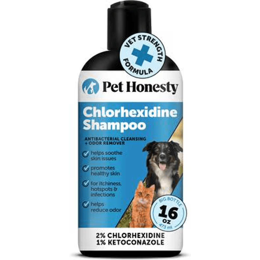
A vet-strength medicated shampoo with 2% chlorhexidine and 1% ketoconazole, the antifungal and antibacterial combination vets use to help clear ringworm and skin infections and to cut fungal spore shedding during treatment.
Webvet may earn a commission when you click through to Chewy, at no extra cost to you.
The pattern that should trigger a biopsy conversation:
- Itchy, scaly, red skin in a dog 8 years or older with no prior allergy history
- Improvement on steroids that never lasts once the dose drops
- Skin that keeps worsening through two or more failed treatment trials
- Depigmentation or sores at the lips, nose, eyelids, or other mucocutaneous junctions
Cutaneous vs. Systemic Lymphoma in Dogs
Cutaneous lymphoma is one branch of a larger disease family, and the two main forms behave very differently. Systemic (multicentric) lymphoma is far more common, typically announces itself with enlarged lymph nodes under the jaw and behind the knees, and is usually a B-cell cancer treated with multi-drug injectable chemotherapy protocols. Cutaneous lymphoma lives in the skin, is almost always T-cell, and is treated primarily with oral chemotherapy. Staging, drug choices, and expected outcomes differ enough that the two are essentially separate diagnoses. For the full picture of the more common systemic form, including symptoms, causes, and staging, see our complete guide to lymphoma in dogs.
How Vets Diagnose Cutaneous Lymphoma
The only definitive diagnosis is a skin biopsy read by a pathologist. Everything else is supporting evidence.
A typical diagnostic path looks like this:
- Fine needle aspiration (FNA). If nodules or enlarged lymph nodes are present, a needle sample may reveal abnormal lymphocytes in minutes. FNA is quick and cheap, but it frequently misses early-stage disease.
- Skin biopsy. Under sedation or brief anesthesia, the vet removes several small punches of affected skin. The pathologist looks for malignant T-cells infiltrating the epidermis and the Pautrier microabscesses that confirm epitheliotropic lymphoma.
- Immunohistochemistry and clonality (PARR) testing. When biopsy findings are ambiguous, these lab tests confirm the cells are T-cells and that they are a malignant clone rather than an inflammatory reaction.
- Staging. Once lymphoma is confirmed, bloodwork, urinalysis, chest X-rays or abdominal ultrasound, and lymph node sampling check whether the disease has moved beyond the skin.

Skin relief and repair capsules that target the gut-skin axis, supporting the skin barrier and easing itch and allergy symptoms in cats and dogs.
Webvet may earn a commission when you click through to Animal Biome, at no extra cost to you.
One practical note: steroids can shrink lymphoma cells enough to blur biopsy results. If your dog is on prednisone and a biopsy is planned, your vet may ask you to pause the medication first. Do not stop any medication without your vet's instruction, but do mention every drug your dog is receiving before the biopsy.
Diagnosis is also the right moment to ask for a referral to a veterinary oncologist. Cutaneous lymphoma is rare enough that most general practitioners see only a handful of cases in a career, while oncologists manage it routinely and have access to current protocols and clinical trials, as the American College of Veterinary Internal Medicine notes in its specialty guidance.
Treatment Options and Response Rates
There is no reliable cure for canine cutaneous lymphoma. Treatment aims to shrink lesions, control itch and pain, and buy good-quality months. Within that honest frame, treatment genuinely helps most dogs feel better.
- Lomustine (CCNU) is the standard first-line chemotherapy. It is an oral capsule given at home every three weeks, with regular bloodwork to monitor liver values and white cell counts. In the largest published study, Risbon and colleagues (Journal of Veterinary Internal Medicine, 2006) reported an overall response rate of about 78 percent in dogs with cutaneous epitheliotropic lymphoma, with roughly one in six achieving complete remission. Responses averaged around three months before the disease returned.
- Prednisone is used alone when chemotherapy is declined, or alongside lomustine. It reduces inflammation and itch and can shrink lesions, but responses are partial and typically brief.
- Radiation therapy can control localized disease, particularly single oral or solitary skin lesions.
- Safflower oil, retinoids, and interferon appear in the literature as adjunct options with modest, inconsistent benefit; a veterinary oncologist can say whether any fit your dog's case.
- Skin-support care matters daily: a chlorhexidine shampoo for secondary infections, antibiotics when ulcerated lesions become infected, and pain or itch control drugs such as gabapentin or oclacitinib as directed by your vet. Daily omega-3 fatty acids support the skin barrier alongside treatment (confirm dosing with your vet), and a lightweight recovery suit can shield ulcerated patches from scratching between baths.
What is the survival rate for dogs with cutaneous lymphoma? Framed as response rather than cure: about three-quarters of treated dogs improve meaningfully on lomustine, but nearly all eventually relapse, and long-term survival beyond a year is uncommon regardless of protocol. There is no published five-year survival rate because durable cures are rare. The realistic goal of treatment is months of comfortable, good-quality life, and by that measure treatment succeeds for most dogs who receive it.
Prognosis: Life Expectancy With Cutaneous Lymphoma
Honesty matters most here, so here are the published numbers rather than soft reassurance.
What is the life expectancy of a dog with cutaneous lymphoma? Most studies place median survival around six months from diagnosis, with wide survival ranges on both sides. Dogs treated with lomustine-based chemotherapy commonly live in the range of six to nine months, while dogs managed with comfort care typically get a few months with prednisone alone, often two to four. A minority of dogs with slowly progressive, early-stage disease live well past a year, and rare indolent cases persist for two years or more, while advanced nodular or Sezary-type disease can shorten survival to weeks.
| Treatment path | Typical survival | What it looks like day to day |
|---|---|---|
| Lomustine (CCNU) chemotherapy | About 6 to 9 months median | Oral capsules every 3 weeks, bloodwork monitoring, good quality of life during response |
| Prednisone and comfort care | About 2 to 4 months | Reduced itch and inflammation, minimal vet visits, gradual progression |
| Early-stage indolent disease | Sometimes 12 months or more | Slow progression, treatment adjusted as lesions advance |
| Advanced nodular or Sezary-type disease | Weeks to a few months | Focus shifts to comfort, wound care, and quality-of-life monitoring |
Owners also ask how quickly does cutaneous lymphoma spread once it is diagnosed. Prognosis and pace are linked: indolent patch-stage disease can smolder for a year or more, while aggressive cases move from plaques to ulcerated nodules, and sometimes to lymph nodes and internal organs, within a few months. Your vet may recheck nodules with a fine needle aspiration to catch that spread early.
Factors that tilt the prognosis:
- Stage at diagnosis. Patch-stage dogs do meaningfully better than dogs diagnosed with ulcerated nodules, which is the entire argument for early biopsy.
- Extent of disease. Solitary lesions treated with surgery or radiation occasionally do very well; generalized disease does not.
- Blood involvement. Sezary syndrome and bone marrow involvement carry the shortest survival times.
- Response to the first protocol. Dogs who achieve complete remission on lomustine hold their gains longer than partial responders.
Because survival is measured in months, quality of life becomes the daily yardstick. Track appetite, sleep, itching, sore ulcerated spots, and enthusiasm for walks, and agree with your vet in advance on which changes would signal that comfort is slipping. That plan spares you from making the hardest decisions in a crisis.
How Long Do Dogs Live With Lymphoma Overall?
Owners researching a new diagnosis often see survival numbers for lymphoma in general and assume they apply to the skin form, but the general figures mostly describe systemic lymphoma, which follows different math. Untreated multicentric lymphoma typically allows one to two months, while dogs treated with multi-agent CHOP chemotherapy commonly reach twelve months or more, numbers that do not transfer to cutaneous disease. For the full breakdown of survival by lymphoma type, stage, and treatment choice, see our dedicated guide to dog lymphoma life expectancy.
Cutaneous Lymphoma FAQs
Our sister site Petful covers the neighboring topic in skin cancer in cats and dogs, useful context for how vets think about skin malignancies overall.
Frequently Asked Questions
Is cutaneous lymphoma painful or itchy for dogs?
Yes, usually both. Intense itching is a hallmark of the disease from the earliest patch stage, and ulcerated plaques and nodules in later stages are genuinely painful, especially when secondarily infected. This is why a comfort care plan matters as much as chemotherapy: itch control medication, pain relief, and prompt treatment of infected sores keep dogs comfortable through every stage. Tell your vet immediately if your dog seems sore, licks lesions constantly, or withdraws from touch, because comfort can almost always be improved with adjusted medication.
Is cutaneous lymphoma in dogs contagious to other pets or people?
No. Cutaneous lymphoma is not contagious to other dogs, cats, or humans in any form of contact. It is a cancer of the dog's own lymphocytes, not an infection, so there is nothing to transmit through touching, grooming, shared bedding, or licking. You do not need to isolate your dog or restrict contact with family members. The only household precaution involves chemotherapy handling: wear gloves when giving lomustine capsules and follow your vet's guidance on cleaning up urine or feces for a few days after each dose.
When to See Your Vet
Cutaneous lymphoma rewards early suspicion more than any other skin cancer in dogs, because the biopsy that diagnoses it is simple and the disease is far more manageable at the patch stage than the nodule stage.
Book an appointment, and raise the biopsy question directly, if your dog has:
- Red, scaly, itchy skin that has failed two or more rounds of allergy treatment
- Skin problems that improve on steroids but always relapse
- New depigmentation or sores on the nose, lips, eyelids, or gums
- Raised plaques or nodules forming in previously flat irritated skin
Most itchy dogs have allergies, and most will be fine. But the small number with cutaneous lymphoma in dogs lose their best treatment window to months of well-intentioned allergy trials. One biopsy settles the question, and asking for it is the single most useful thing an owner in this situation can do.

Editor
The Webvet Editorial Team is the in-house group of pet-care editors and writers behind Webvet, operated by Smart Pet Collective. The team researches, writes, and maintains Webvet's pet health, behavior, and medication content. Every article follows a defined editorial process: research from reputable veterinary and scientific sources, careful drafting, mandatory review of medical content by a credentialed veterinarian, and dated publication. Health and medication articles are medically reviewed by a licensed veterinary professional before they go live and are kept current over time.
Veterinarian · BVMS MRCVS
Dr. Pippa Elliott, BVMS, MRCVS, is a veterinarian with nearly 30 years of experience in companion animal practice. Dr. Elliott earned her Bachelor of Veterinary Medicine and Surgery from the University of Glasgow. She was also designated a Member of the Royal College of Veterinary Surgeons. Married with 2 grown-up kids, Dr. Elliott has a naughty Puggle named Poggle, 3 cats and a bearded dragon.


