Mast Cell Tumor in Dogs: Grades, Treatment, and What to Expect
Most mast cell tumors in dogs are low grade and curable with surgery, but grade decides everything. This vet-reviewed guide puts grades and stages in one table, explains FNA-first diagnosis, and covers treatment, costs, and recurrence.

BVMS MRCVS
Medically reviewed by Dr. Pippa Elliott, BVMS MRCVS · Last reviewed

This article contains affiliate links. Webvet may earn a commission when you buy through them, at no extra cost to you.
A mast cell tumor in dogs is the diagnosis most owners hear right after a lump they barely worried about turns out to matter. Mast cell tumors are the most common malignant skin tumor in dogs, accounting for roughly 16 to 21 percent of all canine skin tumors according to VCA Animal Hospitals. That sounds frightening, but here is the honest version: most mast cell tumors are low grade, most low-grade tumors are cured with one well-planned surgery, and the single biggest factor in what happens next is not the lump's size or how scary it looks. It is the grade your pathologist assigns after testing.
This guide walks you through the whole decision path: what a mast cell tumor is, what it looks and feels like, how grades and stages differ (in one table, because no one explains this clearly), how diagnosis works, why your vet reaches for Benadryl, treatment by grade, real cost ranges, recurrence, and which breeds carry the most risk.

 Sponsored
Want a vet's take on your dog's skin or coat?
Talk to a licensed vet at Dutch over video, often the same day. They can recommend treatment and prescribe it when it's appropriate.
Use code Pets30
Plans start at $11/month · covers up to 5 pets
- Licensed vets in most states
- Rx for 150+ conditions
- 4.7 on Trustpilot
Why Vets Call the Mast Cell Tumor the Great Pretender
- 1Mast cell tumors are cancer, but most are low grade and curable with surgery alone.
- 2Grade, determined by a pathologist, decides nearly everything about treatment and outlook.
- 3Never squeeze or repeatedly handle a suspected mast cell tumor; it can release histamine.
- 4The next step for any new lump is a fine needle aspirate (FNA), a quick needle sample done awake.
So what is a mast cell tumor in dogs, exactly? Mast cells are normal immune cells that live in the skin and other tissues. Their job is to release histamine and other inflammatory chemicals during allergic reactions. A mast cell tumor forms when these cells replicate out of control, creating a mass packed with histamine-loaded granules.
Yes, it is cancer. Mast cell tumors are malignant, which means they have the potential to invade surrounding tissue and spread. But malignancy is a spectrum, not a verdict. VCA Animal Hospitals notes that the majority of mast cell tumors are low grade, and low-grade tumors that are completely removed are usually cured.
Veterinarians call the mast cell tumor "the great pretender" because it can look like almost anything:
- A soft, squishy lump that feels exactly like a harmless fatty lipoma
- A pink, raised button that mimics a benign histiocytoma
- A red, angry, ulcerated sore
- A lump under the skin with no surface change at all
That disguise is exactly why no vet, no matter how experienced, can diagnose a mast cell tumor by look or feel alone. Testing is not optional. If you have just found a new lump and have not seen a vet yet, start with our lump on your dog triage guide to work through the first steps.

Sponsored Lemonade pet insurance link. Webvet may earn a commission.
Symptoms: What a Mast Cell Tumor Looks and Feels Like
The first signs of a mast cell tumor are usually visual: a new lump or bump on or under the skin that was not there before, most often on the trunk, limbs, or head. What makes mast cell tumors distinctive is not one appearance but their behavior over time. A raised red lesion that changes size from week to week, or even day to day, is a classic mast cell presentation, because the tumor swells when its cells release histamine and shrinks as the inflammation settles.

Watch for these patterns:
- A lump that changes size. Growing, shrinking, then growing again is a red flag specific to mast cell tumors.
- Redness and swelling around the mass, sometimes appearing suddenly after the dog scratches or bumps it.
- Itchiness. Dogs often lick or chew at mast cell tumors because histamine makes them itch.
- Ulceration. The surface may break open, weep, or bleed.
- Hives or flushing near the mass after it is handled.
- Darier's sign. Stroking or bumping the lump makes it visibly redden and swell within minutes. This wheal-and-flare reaction is caused by localized histamine release and is so characteristic that vets treat it as a strong clue on its own.
Sudden whole-body signs matter too. A dog with a large or actively degranulating tumor can show vomiting, poor appetite, dark tarry stool, or lethargy, all driven by histamine hitting the stomach. Any of those alongside a skin lump moves the vet visit from this week to today.
Location matters too. Mast cell tumors appear anywhere, but some sites carry different implications:
- Leg and trunk tumors are the most common and often the most straightforward to remove.
- Mouth and muzzle tumors tend to behave more aggressively and spread to lymph nodes earlier, according to the Merck Veterinary Manual.
- Ear, eyelid, and paw tumors are challenging because there is little spare skin for wide surgical margins.
- Subcutaneous tumors sit under the skin rather than in it, feel like soft moveable lumps, and as a group carry a better outlook than skin-based tumors of similar grade.
- Internal mast cell tumors (spleen, liver, intestine) are uncommon in dogs, more serious, and usually found during workup of a sick dog rather than as a lump.
Because photographs beat descriptions for this disease, we keep a dedicated library of real mast cell tumor pictures showing low-grade, high-grade, ulcerated, and subcutaneous presentations so you can compare your dog's lump before the vet visit. And if the lump on your dog is a smooth, fluid-filled swelling rather than a firm mass, review our guide to the dog cyst, a common benign look-alike.

A collar-mounted sensor and app for activity, rest, sleep, respiratory rate, heart-rate trends, itch behaviors, and more.
- Builds a personalized baseline over time
- Tracks trends between veterinary visits
- Generates a month-long shareable report
Health monitoring for dogs and cats
Webvet may earn a commission when you click through to Maven Pet, at no extra cost to you.
Grades vs. Stages, Explained in One Table
Here is the concept that confuses nearly every owner, because most articles explain grading and staging in separate sections and never connect them. They answer two different questions:
- Grade asks: how aggressive are the tumor cells themselves? It is the biopsy answer, assigned by a pathologist looking at tissue under a microscope after removal.
- Stage asks: how far has the tumor spread through the body? It is the imaging-and-sampling answer, based on lymph node testing, ultrasound, and sometimes bone marrow checks.
Pathologists actually use two grading systems side by side. The older Patnaik system (published by Dr. Amiya Patnaik and colleagues in 1984) assigns grade 1, 2, or 3. Because the middle grade 2 category was frustratingly unpredictable, Michigan State University pathologist Dr. Matti Kiupel and colleagues published a simpler two-tier system in 2011 that classifies every tumor as either low grade or high grade. Most pathology reports today include both. Staging follows the World Health Organization's 0 to 4 scheme.
Most explanations cover Patnaik grade, Kiupel grade, and clinical stage in separate sections and leave you to connect them yourself. So here is the single combined grades-and-stages table in one owner-readable chart: all three scales together, crossing Patnaik grades 1 to 3 and the Kiupel high/low call with clinical stage 0 to 4, so you can find your dog's pathology results on one chart:
| Scale | Value | What it means | Typical implication for treatment |
|---|---|---|---|
| Patnaik grade | Grade 1 | Well-differentiated cells confined to the skin | Surgery alone is usually curative |
| Patnaik grade | Grade 2 | Intermediate cells; behavior hard to predict | Surgery with wide margins; Kiupel grade breaks the tie |
| Patnaik grade | Grade 3 | Poorly differentiated, aggressive cells | Surgery plus oncology referral for chemo or Palladia |
| Kiupel grade | Low grade | Predicted to behave benignly after removal | Complete excision, then monitoring |
| Kiupel grade | High grade | Predicted to spread and recur | Full staging plus systemic therapy with an oncologist |
| Clinical stage | Stage 0 | Incompletely removed tumor, no node spread | Second surgery or radiation to clean up margins |
| Clinical stage | Stage 1 | One skin tumor, no lymph node involvement | Local treatment (surgery) is usually enough |
| Clinical stage | Stage 2 | One tumor with regional lymph node spread | Surgery plus node removal and systemic therapy |
| Clinical stage | Stage 3 | Multiple large or deep tumors, with or without nodes | Combination therapy planned by an oncologist |
| Clinical stage | Stage 4 | Distant spread (spleen, liver, blood, marrow) | Systemic therapy focused on comfort and control |
Read your dog's pathology report against this chart and the plan starts to make sense. A Patnaik grade 2, Kiupel low-grade, stage 1 tumor is a surgery-and-monitor situation. A Kiupel high-grade tumor at any stage is an oncologist conversation. A "stage 2 mast cell tumor" is not automatically dire; it means the local lymph node is involved and the plan expands to address it.
Two clarifications the table cannot carry on its own. A subcutaneous mast cell tumor in dogs, one that sits under the skin rather than in it, is graded on the same systems but tends toward better behavior than a skin tumor of matching grade. And treatment for a low grade mast cell tumor in dogs is usually the simplest line on the chart: complete surgical removal, then monitoring.
One practical note on grade 2 tumors, since they are the most common and the most confusing: when the Patnaik system says "intermediate," ask your vet what the Kiupel grade is. Low-grade behavior is the norm; the Kiupel result tells you whether your dog's tumor is the exception. For what each grade means for lifespan, our companion guide to mast cell tumor life expectancy by grade walks through the published survival data in detail.
 Sponsored
Vet care that's always there
Unlimited video calls and messaging with licensed vets for less than a typical vet visit. One membership covers up to five pets.
Use code Pets30
Plans start at $11/month · covers up to 5 pets
- Licensed vets in most states
- Rx for 150+ conditions
- 4.7 on Trustpilot
Diagnosis: FNA First, and Why Your Vet Gives Benadryl
The FNA-first diagnostic path for a suspected mast cell tumor is refreshingly simple, and it comes with one hard rule owners must know before the appointment: never squeeze the lump. FNA is a quick procedure done in the exam room, awake, no sedation: the vet inserts a thin needle into the lump, draws out a sample of cells, and examines them under a microscope. Mast cells are one of the easiest cell types to identify because their histamine granules stain a distinctive purple. Many vets can confirm a mast cell tumor in-house within minutes; ambiguous samples go to a clinical pathologist.
For a focused medication reference, see WebVet’s vet-reviewed Famotidine for Dogs and Cats (Pepcid): Dosage Chart and Safety guide, including the dosage, side effects, and safety questions to confirm with your veterinarian.
For a focused medication reference, see WebVet’s vet-reviewed Benadryl for Dogs: Dosage Chart by Weight and Safety Guide guide, including the dosage, side effects, and safety questions to confirm with your veterinarian.

This is why the FNA-first rule matters so much: the great pretender problem cuts both ways. A histiocytoma, a benign immune tumor common in young dogs, can look identical to a mast cell tumor by eye, and the two demand completely different responses. One resolves on its own; the other needs planned surgery. Our guide to histiocytoma, the benign look-alike covers how vets tell them apart. The needle answers in minutes what the eye cannot answer at all.
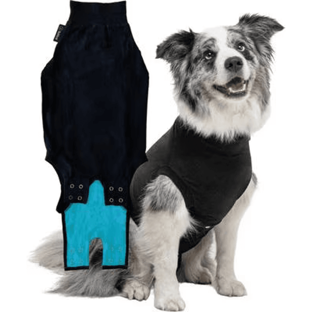
A breathable full-body recovery suit that covers a spay or abdominal incision, the vet-recommended cone alternative that lets a dog rest, walk, and go potty normally without licking or scratching the wound.
Webvet may earn a commission when you click through to Chewy, at no extra cost to you.
Now the question owners ask constantly: why do vets give Benadryl around diagnosis and surgery for a mast cell tumor in dogs? It is not a treatment, and it does not shrink the tumor. Your vet pre-treats with Benadryl as a shield against histamine release, and the logic deserves its own plain-language explanation: pre-treating blunts degranulation before the needle or scalpel can provoke it. Every mast cell tumor is a warehouse of histamine, and anything that disturbs the tumor, an FNA, surgical handling, even everyday friction, can release some of it. Diphenhydramine (Benadryl) is an antihistamine that blocks H1 receptors, so the released histamine has fewer places to act.
Vets deploy it at specific decision points:
- Before or after an FNA on a reactive-looking tumor, to blunt the local swelling the needle can provoke.
- In the days before surgery, so histamine released during handling of the tumor does not cause dangerous drops in blood pressure under anesthesia.
- While waiting for surgery, often paired with a stomach protectant like famotidine, because chronic histamine release drives stomach acid and can ulcerate the gut.
Never dose Benadryl on your own for a tumor; the timing and combination with acid blockers is a medical decision. But when your vet prescribes it, this is why: the drug is managing the tumor's chemistry, not the tumor itself.
If the FNA confirms mast cells, your vet may recommend staging before surgery, typically an aspirate of the nearest lymph node, and for higher-risk tumors an abdominal ultrasound to check the spleen and liver. For most small, low-risk skin tumors, vets proceed to surgery and let the biopsy grade guide everything after.
Treatment by Grade: Surgery, Stelfonta, Radiation, and Palladia
Should you remove your dog's mast cell tumor? In almost every case where surgery is feasible, yes, and sooner rather than later. Small tumors allow clean wide margins, completely excised low-grade tumors are usually cured outright, and waiting only makes the surgery bigger and gives an aggressive tumor time to spread. The exceptions are locations where removal would be disfiguring and dogs for whom anesthesia is the greater risk; those cases are exactly what the non-surgical options below exist for.
Beyond that decision, treatment for a mast cell tumor in dogs is not one-size-fits-all; it is a menu, and grade picks from it. Here is how the options map to the situation.
- Surgery is the foundation for nearly every mast cell tumor. Because microscopic tumor cells extend beyond the visible lump, surgeons take wide margins: the standard is roughly 2 centimeters of normal-looking skin on all sides plus one tissue layer below, per the Merck Veterinary Manual. That is why the incision is always longer than the lump seemed to justify. The pathologist then checks whether the margins are "clean" (no tumor cells at the cut edge). For Patnaik grade 1 and Kiupel low-grade tumors, complete excision with clean margins is usually the end of the story.
- Stelfonta (tigilanol tiglate) is the injection option. Approved by the FDA in 2020, Stelfonta is a drug injected directly into the tumor by a veterinarian, where it destroys the tumor tissue, which then sloughs away as an open wound that heals over several weeks. In the pivotal clinical study submitted for FDA approval, a single injection achieved complete remission in 75 percent of treated tumors. It is an option for non-metastatic skin tumors up to about 8 cubic centimeters in spots where surgery is difficult, such as the lower leg. To be clear for anyone searching "how to shrink a mast cell tumor at home": this is a vet-administered prescription procedure with mandatory pre-medication (including antihistamines and steroids, for exactly the degranulation reasons above). There is no home version, and no supplement or natural remedy has been shown in any published veterinary study to shrink a mast cell tumor. Delaying real treatment to try one gives the tumor time to grow beyond easy removal.

A lightweight, padded fabric cone that gently blocks a pet from pawing, scratching, or rubbing a healing eye, wound, or hot spot, and it is far softer and less stressful than a hard plastic cone.
Webvet may earn a commission when you click through to Chewy, at no extra cost to you.
- Radiation therapy cleans up what surgery cannot. When margins come back incomplete and a second surgery is not possible, radiation to the surgical site offers excellent long-term control for low and intermediate grade tumors; VCA Animal Hospitals describes control rates in this setting as among the best in veterinary radiation oncology.
- Palladia (toceranib phosphate) is the pill for high-grade and spread disease. Palladia was the first drug FDA-approved specifically to treat cancer in dogs (2009), and it targets the KIT receptor pathway that drives many aggressive mast cell tumors. In its pivotal study, about 43 percent of dogs with recurrent or spread mast cell tumors had an objective response. It is prescribed and monitored by a veterinarian, usually an oncologist, with regular blood work.
- Traditional chemotherapy (vinblastine with prednisone) remains a standard oncologist-directed protocol for high-grade tumors after surgery, and prednisone alone is sometimes used to shrink a tumor before surgery to make margins achievable. Chemotherapy in dogs is dosed for quality of life, not the punishing intensity people associate with human oncology; most dogs sail through with mild or no side effects, and the protocol stops if it costs more comfort than it buys. GI upset is the side effect owners manage most at home, so ask your oncologist about adding a veterinary probiotic such as Purina FortiFlora during treatment.
For a focused medication reference, see WebVet’s vet-reviewed Omeprazole for Dogs and Cats: Dosage Chart, Side Effects, and Cost guide, including the dosage, side effects, and safety questions to confirm with your veterinarian.
Whatever the plan, ask your vet three questions before treatment starts. Those three answers define the entire road ahead.
- What is the Kiupel grade?
- Are clean margins realistic at this location?
- Does anything on staging change the goal from cure to control?
Matching plan to grade:
- Kiupel low grade / Patnaik 1: surgery with clean margins, then monitoring. Stelfonta where surgery is awkward.
- Patnaik 2, Kiupel low: surgery with wide margins; radiation or a second surgery if margins are dirty.
- Kiupel high grade / Patnaik 3: surgery plus full staging plus systemic therapy (vinblastine protocol or Palladia) with a veterinary oncologist.
- Stage 2 and up (node or distant spread): node removal where feasible plus systemic therapy; goals shift toward long-term control and quality of life. Comfort gear earns its keep here: an orthopedic bed cushions pressure points as activity drops.
Removal Costs, Recovery, and Recurrence
Cost is the question almost no veterinary source answers in writing. So here is a cost table by treatment path, with realistic U.S. ranges. Your market, your dog's size, and general practice versus specialty pricing move these numbers substantially; treat them as planning figures, not quotes, and ask your own clinic for a written estimate before scheduling anything.
| Treatment path | Typical U.S. cost range | What it covers |
|---|---|---|
| Fine needle aspirate + cytology | $100 - $400 | Needle sample and pathologist review |
| Staging workup | $300 - $1,500 | Lymph node aspirate, blood work, abdominal ultrasound |
| Surgical removal (general practice) | $500 - $2,000 | Anesthesia, wide-margin excision, biopsy with grading |
| Surgical removal (board-certified surgeon) | $2,000 - $5,000 | Complex sites, reconstruction, difficult margins |
| Stelfonta injection | $1,000 - $2,500 per treatment | Drug, pre-medication protocol, wound rechecks |
| Radiation therapy course | $4,500 - $10,000 | Planning plus 15 - 20 treatment sessions |
| Palladia | $300 - $800 per month | Drug plus monitoring blood work |
| Chemotherapy (vinblastine protocol) | $2,000 - $4,000 per course | Drugs, administration, monitoring |
A few cost realities worth naming:
- The FNA is the cheapest decision-quality money you will spend. A $150 needle sample is what tells you whether you are facing a $700 problem or a $7,000 one.
- Small tumors are cheap tumors. The single most effective cost-control move is acting while the lump is small enough for easy wide margins.
- Pet insurance typically covers mast cell tumors diagnosed after enrollment; a pre-existing tumor will not be covered, which is another argument against watchful waiting on an untested lump.

Recovery from routine removal is usually 10 to 14 days. Expect:
- An incision noticeably longer than the lump seemed to justify (that is the wide margin, not an error)
- A soft cone or recovery suit full time until sutures come out
- Leash-only activity restriction, since stretching the incision line risks opening it
- Mild bruising or swelling for the first few days, sometimes managed with a short antihistamine course
- Suture removal and margin report review at the two-week recheck
Stelfonta recovery looks different and alarming if you are not warned: the tumor site opens into a wound as the dead tissue sloughs, then granulates and closes over four to six weeks. That wound is the treatment working as designed.
Recurrence is where grade and margins earn their importance. Completely excised low-grade tumors rarely come back at the surgery site; incompletely excised tumors can, which is why dirty margins trigger a second surgery or radiation rather than a shrug. The bigger long-term issue is new, unrelated mast cell tumors: VCA Animal Hospitals notes that dogs that have developed one mast cell tumor are at increased risk of developing others, and the Merck Veterinary Manual reports that dogs may develop multiple tumors over their lifetime. That does not mean spread, and each new tumor gets its own FNA and its own grade.
Because that recurrence risk is lifelong, recurrence risk and the recheck schedule belong in the same conversation with your vet before you leave the post-surgery recheck. The schedule below is the standard surveillance rhythm most vets recommend after a mast cell tumor, and it stays in place for the rest of your dog's life, tightening only if a high grade or dirty margins raise the risk:
- Monthly home lump checks for life: hands over the whole dog, note anything new on a phone photo with a coin for scale.
- Vet recheck of the surgical site and local lymph node every 3 months for the first year, then every 6 months.
- Immediate FNA of any new lump, no matter how harmless it feels. Never assume the new bump is "just another one like last time."
What Causes Mast Cell Tumors and Which Breeds Are at Risk
So what triggers mast cell tumors in dogs? The honest answer is that no owner-controllable cause has been identified. You did not feed the wrong food, use the wrong flea product, or miss a warning sign that would have prevented this. The best-understood driver is genetic: mutations in the c-KIT gene, which codes for a growth receptor on mast cells, are found in roughly a quarter to a third of intermediate and high-grade tumors, according to the Merck Veterinary Manual. That KIT pathway is exactly what Palladia targets, and your pathologist can run KIT testing on the biopsy to help predict behavior and drug response. Chronic skin inflammation has been proposed as a contributing factor, but the evidence is associative, not causal.
The breed pattern is the strongest clue that genetics leads, and breed risk concentrates heavily in Boxers and other brachycephalic breeds, the short-nosed dogs whose shared ancestry seems to carry the predisposition. Knowing your dog's breed risk changes nothing about prevention, but it should change how seriously you take every new lump, including how fast it gets a needle. Higher-risk breeds include:
- Boxers, the poster breed for this disease, along with Boston Terriers, Pugs, and English Bulldogs
- Golden Retrievers and Labrador Retrievers
- Shar-Peis, notable because their tumors skew aggressive and appear at younger ages
- Rhodesian Ridgebacks, Weimaraners, and Beagles
Here is the reassuring wrinkle in the Boxer story: VCA Animal Hospitals notes that while Boxers and related brachycephalic breeds develop mast cell tumors far more often than average, their tumors are disproportionately low grade. A mast cell tumor in a Boxer is a common event with a usually favorable script. A Shar-Pei's tumor deserves more immediate concern.
Age-wise, this is mostly a middle-aged and senior dog disease, with the Merck Veterinary Manual citing a mean age around 9 years. Mast cell tumors in young dogs do occur, and a lump on a 2-year-old still deserves an FNA, but statistically a young dog's pink button lump is more likely a histiocytoma. There is no meaningful sex predisposition, and no evidence that spaying or neutering changes risk in a way that should drive decisions.
There is also no prevention. The only lever you control is surveillance: monthly hands-on lump checks and an FNA-first response to anything new. Owners of high-risk breeds should treat that routine as seriously as heartworm prevention.
See Real Mast Cell Tumor Pictures
Because this tumor impersonates so many harmless lumps, mast cell tumor pictures in dogs are worth a thousand words of description. We keep a dedicated visual library of real mast cell tumor in dogs pictures showing low-grade, high-grade, ulcerated, and subcutaneous presentations, each labeled with what to notice. If you are trying to match your dog's lump against a picture of a mast cell tumor in dogs before the vet visit, start there, and remember that no photo comparison replaces the FNA.
Prognosis Snapshot: Life Expectancy by Grade
Because grade dominates outcome, the prognosis question deserves its own honest treatment, and we give it one in a dedicated companion article. The short version: in the Kiupel two-tier study of 95 dogs (Kiupel et al., Veterinary Pathology, 2011), dogs with high-grade tumors had a median survival of less than 4 months, while dogs with low-grade tumors had a median survival beyond the study's 2-year follow-up window. Patnaik's original 1984 study reported roughly 93 percent of grade 1 dogs alive at 1,500 days versus 6 percent of grade 3 dogs. Those numbers are group statistics, not your dog's sentence, and they shift meaningfully with margins, location, stage, and treatment choices. For the full grade-by-grade and stage-by-stage breakdown, including what changes the odds, see our complete guide to mast cell tumor life expectancy by grade.
For a focused medication reference, see WebVet’s vet-reviewed Sucralfate for Dogs: Dosage, Timing, and How to Give It guide, including the dosage, side effects, and safety questions to confirm with your veterinarian.
Mast Cell Tumor FAQs
If you want a shorter second read after this one, our sister site Petful keeps a quick-reference guide to mast cell tumors in dogs.
Frequently Asked Questions
Should I remove my dog's mast cell tumor?
Yes, you should remove your dog's mast cell tumor in almost every case where surgery is feasible, and sooner rather than later. Grade and location decide the details, but the math favors early removal of a mast cell tumor: small tumors allow clean wide margins, and completely excised low-grade tumors are usually cured outright. Waiting lets the tumor grow into a harder, more expensive surgery and gives an aggressive tumor time to spread. The exceptions are tumors in locations where surgery would be disfiguring (where Stelfonta or radiation may serve instead) and dogs whose age or health makes anesthesia the greater risk. Discuss margins with your vet before the procedure, ask whether 2 centimeter margins are achievable at that site, and always submit the removed tissue for grading.
How serious is a mast cell tumor in dogs?
It is a real cancer diagnosis, so it is always serious enough to treat, but the range of outcomes is enormous and the odds favor your dog. Most mast cell tumors are low grade, and most low-grade tumors are cured with complete surgical removal. High-grade tumors are genuinely dangerous, spread readily, and need an oncologist's help, but they are the minority. Until the pathology report comes back, you do not know which situation you are in, which is exactly why the FNA and biopsy matter more than anything you can see or feel from the outside. Treat every mast cell tumor promptly; panic about none of them until grade says otherwise.
Can a dog live a normal life after mast cell tumor removal?
Most dogs do. A dog with a completely removed low-grade tumor typically returns to full normal life after the two-week surgical recovery, with no ongoing medication and no restrictions. What changes permanently is the surveillance: monthly lump checks at home, periodic vet rechecks of the site and nearby lymph node, and a same-week FNA for any new bump, because dogs that have had one mast cell tumor are more likely than average to develop another somewhere else. Dogs on long-term Palladia for high-grade disease can also enjoy good quality of life, with the trade-off of regular monitoring blood work.
What can be mistaken for a mast cell tumor in dogs?
Nearly any lump, in both directions, which is why vets call this tumor the great pretender. Benign masses commonly confused with mast cell tumors include lipomas (soft fatty lumps), histiocytomas (pink button tumors of young dogs), skin tags, warts, and cysts. Meanwhile a mast cell tumor can impersonate every one of those. Insect bites and hives can also mimic the sudden red swelling of a degranulating tumor. No visual inspection settles it; only a fine needle aspirate does. The purple-staining granules inside mast cells make them one of the most recognizable cell types under the microscope, so a quick needle sample usually gives a same-day answer.

Editor
The Webvet Editorial Team is the in-house group of pet-care editors and writers behind Webvet, operated by Smart Pet Collective. The team researches, writes, and maintains Webvet's pet health, behavior, and medication content. Every article follows a defined editorial process: research from reputable veterinary and scientific sources, careful drafting, mandatory review of medical content by a credentialed veterinarian, and dated publication. Health and medication articles are medically reviewed by a licensed veterinary professional before they go live and are kept current over time.
Veterinarian · BVMS MRCVS
Dr. Pippa Elliott, BVMS, MRCVS, is a veterinarian with nearly 30 years of experience in companion animal practice. Dr. Elliott earned her Bachelor of Veterinary Medicine and Surgery from the University of Glasgow. She was also designated a Member of the Royal College of Veterinary Surgeons. Married with 2 grown-up kids, Dr. Elliott has a naughty Puggle named Poggle, 3 cats and a bearded dragon.


