Dog Cyst Guide: Pictures, Types, and When to Worry
Most dog cysts are benign sacs of fluid or debris, but you cannot confirm that by feel. See cyst types with photos, learn how vets rule out tumors with a quick needle test, get burst-cyst first aid steps, and see honest removal cost ranges.

BVMS MRCVS
Medically reviewed by Dr. Pippa Elliott, BVMS MRCVS · Last reviewed

This article contains affiliate links. Webvet may earn a commission when you buy through them, at no extra cost to you.
Finding a dog cyst is one of the most common reasons owners end up combing their pet's skin with a flashlight at midnight. The good news: most cysts on dogs are benign, slow-growing sacs of fluid or debris, not cancer. The catch is that you cannot confirm that by looking or feeling.
This guide shows you what the major cyst types look like with photos, explains how veterinarians actually tell a cyst from a tumor, walks you through first aid if a cyst bursts, and gives honest numbers on removal and cost. If the bump is between your dog's toes, we will point you to the right resource for that specific problem.
- 1Most dog cysts are benign sacs of fluid, sebum, or keratin, but no cyst can be confirmed benign by feel alone.
- 2A fine needle aspiration (FNA) at your vet is the only reliable way to rule out a tumor.
- 3Never squeeze or pop a cyst. If one bursts on its own, clean it gently and call your vet.
- 4Removal is usually elective and typically runs a few hundred dollars, more for tricky locations.

 Sponsored
Want a vet's take on your dog's skin or coat?
Talk to a licensed vet at Dutch over video, often the same day. They can recommend treatment and prescribe it when it's appropriate.
Use code Pets30
Plans start at $11/month · covers up to 5 pets
- Licensed vets in most states
- Rx for 150+ conditions
- 4.7 on Trustpilot
What Is a Dog Cyst?
A cyst is a closed sac in or under the skin, lined by a membrane and filled with fluid, sebum (skin oil), keratin (the protein in hair and nails), or cellular debris. The lining keeps producing material, which is why many cysts slowly enlarge and why drained cysts often refill.
Cysts form when something blocks a normal skin structure. A plugged sebaceous gland traps oil. A blocked or damaged hair follicle traps keratin. Trauma, pressure, friction, and breed-linked skin traits can all set one off, which is why cysts often show up on pressure points and in middle-aged and older dogs.
Three features make a lump behave like a classic cyst:
- It grows slowly over weeks to months, not days.
- It feels round, smooth, and somewhat movable under the skin.
- It may develop a visible pore, a bluish or whitish tint, or discharge a pasty, cottage-cheese-like material.
Beyond the two headline types covered below, dogs can also develop rarer true cysts. Apocrine (sweat gland) cysts are thin-walled, bluish, fluid-filled sacs. Dermoid cysts are congenital pockets of skin tissue, seen along the spine in breeds like the Rhodesian Ridgeback. Your vet will name the exact variety; the owner playbook is the same for all of them.
None of those features prove it is a cyst. Plenty of tumors, including mast cell tumors, can mimic a harmless-looking bump. That is why every new lump earns a vet check, and why this article keeps repeating the FNA rule you will meet below.

Sponsored Lemonade pet insurance link. Webvet may earn a commission.
Types of Dog Cysts, With Photos
Owners searching for a dog cyst almost always want the same thing first: a picture to compare against. This section is a labeled photo gallery for exactly that photo identification job: compare your dog's bump against the sebaceous cyst photo and the follicular cyst photo below, then check the false cysts that get mistaken for both. Match the photo, then still confirm with your vet.
A note on using photos well: compare texture, color, and the presence of a central pore, not just overall shape. Take your own photo in good light before the vet visit, because bumps have a habit of hiding under fur exactly when the vet goes looking. And if what you see is red, ulcerated, or rapidly changing, stop comparing pictures and book the appointment; that presentation has outgrown photo identification.
Sebaceous Cysts

A sebaceous cyst forms when a sebaceous gland or its duct becomes blocked and oil backs up into a sac. On the skin it looks like a round, raised, white or bluish nodule, often 0.5 to 5 centimeters, sometimes with a small dark pore at the center.
If a sebaceous cyst opens, it discharges a thick, grayish-white, pasty material with a greasy feel. Owners often describe it as toothpaste or cottage cheese. That discharge is trapped sebum and keratin, not pus, though a cyst can become secondarily infected and then look red, angry, and painful.
Sebaceous cysts are most common in middle-aged and senior dogs. Cocker Spaniels, Schnauzers, Yorkshire Terriers, and Shih Tzus are among the breeds vets see them in most, according to the clinical overview at VCA Animal Hospitals.
Follicular Cysts

A follicular cyst forms from a hair follicle rather than an oil gland. The follicle's opening gets plugged, and keratin and follicular debris accumulate below it. The result is a firm, dome-shaped bump that sits exactly where a hair shaft should exit the skin.
Follicular cysts feel firmer than sebaceous cysts because their contents are drier and more compacted. When they rupture, the discharge is thick, gray to yellow-brown, and grainy. Ruptured follicular cysts also inflame the surrounding tissue aggressively, because keratin outside its sac triggers a strong foreign-body reaction.
These cysts often appear on pressure points and areas of friction. Some dogs get one; some breeds, including Boxers and Shih Tzus, are prone to crops of them.
Sebaceous Adenomas and Other "False Cysts" That Get Mistaken for Cysts
Several common growths get called cysts by owners but are technically something else:
- Sebaceous adenomas: benign wart-like tumors of the oil glands, very common in older dogs. They look like small pink cauliflower bumps rather than smooth sacs. They are close cousins of dog warts in appearance.
- Lipomas: soft, movable fatty tumors under the skin. Owners often call these "fatty cysts," but a lipoma is a benign fat tumor, not a fluid-filled sac. Our guide to fatty tumors (lipomas) on dogs covers them in depth.
- Histiocytomas: pink, button-like bumps in young dogs that often regress on their own. See our histiocytoma guide.
- Skin tags: soft, dangling flaps of skin, covered in our dog skin tags article.
- Hematomas and seromas: pockets of blood or serum after trauma, sometimes called false cysts because they lack a true cyst lining.
The label matters less than the process: any of these can be confused with a cyst on sight, and a couple of genuinely dangerous tumors can be confused with all of them. That is the problem the next section solves.
Comparing the Common Cyst Types at a Glance
| Type | Typical look | Typical feel | Discharge if opened | Usual concern level |
|---|---|---|---|---|
| Sebaceous cyst | Round white or bluish nodule, may have central pore | Soft to slightly firm, movable | Gray-white, pasty, greasy | Low unless infected or ruptured |
| Follicular cyst | Firm dome at a follicle, often on pressure points | Firm | Thick gray to yellow-brown, grainy | Low, but ruptures inflame badly |
| Sebaceous adenoma | Pink, wart-like, cauliflower surface | Firm, attached | Usually none | Low, cosmetic or irritation |
| "Fatty cyst" (lipoma) | Smooth swelling under normal skin | Soft, doughy, movable | None | Low, monitor size and location |
| False cyst (hematoma or seroma) | Soft swelling after trauma | Fluctuant | Blood or clear fluid if drained | Vet should evaluate cause |

A collar-mounted sensor and app for activity, rest, sleep, respiratory rate, heart-rate trends, itch behaviors, and more.
- Builds a personalized baseline over time
- Tracks trends between veterinary visits
- Generates a month-long shareable report
Health monitoring for dogs and cats
Webvet may earn a commission when you click through to Maven Pet, at no extra cost to you.
Cyst vs Tumor: How to Actually Tell
Here is the answer to the question behind almost every search about a dog cyst: should you be worried? A little, in a productive way. Most cysts are benign, and a bump that has all the classic cyst features usually turns out to be exactly that. But dangerous tumors can imitate cysts closely. Mast cell tumors are notorious shape-shifters that can look like a soft, harmless bump for months. So the right response to any new lump is not panic; it is a vet appointment for a needle sample, ideally within a couple of weeks, sooner if the lump is growing fast, ulcerated, or bothering your dog.
The tool vets use is fine needle aspiration (FNA). Your veterinarian inserts a thin needle into the lump, draws out cells, and examines them under a microscope, often in-clinic during the same visit. FNA takes minutes, rarely needs sedation, and is no more painful than a vaccine.
What the needle finds settles the question the fingers cannot:
- A cyst yields sebum, keratin flakes, and debris with no worrisome cells.
- A lipoma yields fat droplets.
- A mast cell tumor yields distinctive granulated cells that are unmistakable under the microscope.
- An ambiguous sample, or a cell type that needs grading, sends your vet to the next step: a biopsy, where actual tissue goes to a pathologist for the definitive answer.
Why not just watch and wait without the needle? Because the cost of being wrong is asymmetric. If you assume tumor and it is a cyst, you spent a little money on peace of mind. If you assume cyst and it is a mast cell tumor, you gave a malignancy months to grow and possibly spread, and turned a simple excision into a bigger surgery with a worse prognosis. A $25 to $200 test removes that gamble.
- 1No veterinarian, and no owner, can reliably tell a cyst from a tumor by feel. Soft and movable does not mean benign.
- 2Fine needle aspiration (FNA) is the quick, low-cost test that identifies most lumps during a regular visit.
- 3If FNA results are unclear, the next step is a biopsy, which examines actual tissue and gives a definitive diagnosis.
- 4Get every new lump aspirated once, and re-test any lump that changes in size, texture, or color.
A few patterns should raise your urgency level. Book a prompt appointment if the lump:
- appeared suddenly or is growing quickly
- is firm and fixed in place rather than movable
- is ulcerated, bleeding, or changing color
- seems painful, or sits somewhere it interferes with movement, vision, or bathroom habits
- comes with other signs like lethargy, appetite loss, or swollen lymph nodes
If your dog has several mystery bumps and you want a triage framework for all of them, start with our lump on dog triage guide, which walks through the full range from lipomas to lymphoma. This article stays focused on cysts.
One more reassurance backed by data: in studies of canine skin masses, cysts and benign tumors far outnumber malignant ones. The Merck Veterinary Manual's owner resources on skin disorders note that cysts themselves are non-cancerous by definition, though their appearance overlaps with tumors that are not. You can read their overview of canine skin growths at the Merck Veterinary Manual dog owner section. The FNA is what moves your dog's bump from "probably fine" to "confirmed fine."
A painful swelling that develops after a bite or puncture may be an abscess rather than a cyst. See our dog abscess guide for signs of infection and why a burst swelling still needs veterinary assessment.
 Sponsored
Vet care that's always there
Unlimited video calls and messaging with licensed vets for less than a typical vet visit. One membership covers up to five pets.
Use code Pets30
Plans start at $11/month · covers up to 5 pets
- Licensed vets in most states
- Rx for 150+ conditions
- 4.7 on Trustpilot
My Dog's Cyst Burst: First Aid Steps

A ruptured cyst looks alarming: matted fur, an open sore, and thick discharge that may be gray, white, yellow-brown, or blood-tinged. A burst cyst on a dog with blood mixed into the discharge is common and does not automatically mean anything sinister; broken capillaries in the cyst wall bleed easily.
Cysts burst for predictable reasons. The sac fills until pressure splits it, the dog scratches or chews it open, friction from a collar or bedding wears through it, or an infection weakens the wall. Whatever the trigger, your job is the same: protect the wound, do not make it worse, and get the vet involved.
This is the exact moment most advice leaves owners guessing, with nothing more specific than see your vet. So this guide pairs the labeled cyst-type photo gallery above with a concrete burst-cyst first-aid checklist below, built around one hard never-squeeze rule: clean the area gently, cover loosely, and let your veterinarian handle the sac itself.
Here is the first-aid sequence to follow when a cyst has burst:
- Stay calm and restrain gently. Even sweet dogs may snap when a sore spot is handled. Use a helper or a soft muzzle if needed.
- Trim or part the fur around the site if you safely can, so you can see the wound and keep discharge from matting into the coat.
- Clean the area with lukewarm water or saline, or a dilute chlorhexidine solution if you have one in your pet first-aid kit. Wipe discharge away from the wound in outward strokes, using fresh gauze for each pass.
- Do not squeeze the cyst to express remaining material, and skip hydrogen peroxide and rubbing alcohol, which damage healing tissue.
- Cover loosely with a non-stick pad and light wrap if the location allows. The goal is to keep dirt and tongues off, not to compress it.
- Block licking and chewing with an Elizabethan collar or a recovery suit. Licking is the fastest route to infection.
- Call your veterinarian the same day. A ruptured cyst usually needs professional cleaning, often antibiotics, and a plan for the sac itself, because a burst cyst almost always refills or ruptures again until the lining is removed.
The same first-aid checklist applies whatever kind of sac gave way. A sebaceous cyst on a dog popped by scratching drains greasy gray-white material, while a popped follicular cyst on a dog releases drier, grainier debris that inflames the surrounding skin more aggressively; both need the same cleaning, covering, and same-day vet call, and never squeeze either one to finish the job.
Healing timelines are worth knowing so you can judge progress. A cleanly drained, uninfected rupture typically closes over within one to two weeks with basic care. An infected rupture takes longer and often needs a course of oral antibiotics before the skin settles. If the site is still open, weeping, or scabbing and reopening after two weeks, stop waiting: that pattern means the sac is still active underneath, and your vet should reassess.
Owners sometimes ask whether they can just keep cleaning a cyst that bursts every few weeks. You can, but you are treating the drain, not the plumbing. Recurrent rupture is one of the clearest reasons vets recommend removal, covered two sections down.
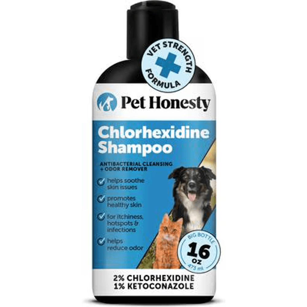
A vet-strength medicated shampoo with 2% chlorhexidine and 1% ketoconazole, the antifungal and antibacterial combination vets use to help clear ringworm and skin infections and to cut fungal spore shedding during treatment.
Webvet may earn a commission when you click through to Chewy, at no extra cost to you.
How Vets Treat Cysts on Dogs
How do you treat cysts on a dog? The honest answer: usually you monitor them, sometimes you medicate them, and you remove them surgically when they cause trouble. There is no cream, supplement, or home remedy that dissolves a true cyst, because the sac lining persists and keeps producing material. Treatment is a staged decision your vet makes with you, based on what the FNA showed, where the cyst is, and how much it bothers the dog.
Can a dog cyst heal on its own? Rarely in any lasting way. A cyst may drain, flatten, or stop growing for a while, but the sac lining stays behind and keeps producing material, so most cysts persist or refill rather than truly healing on their own. That is why treating a cyst on a dog, whether it is a sebaceous or follicular type, follows a ladder that only ends permanently at surgical removal of the lining.
The typical progression looks like this:
- Confirm first. FNA establishes that the lump really is a cyst. Everything after depends on this step.
- Monitor. For a confirmed, small, closed, non-bothersome cyst, benign neglect is legitimate medicine. Measure it monthly, photograph it beside a coin for scale, and recheck if it changes.
- Treat infection. An inflamed or infected cyst gets topical or oral antibiotics and anti-inflammatories. This calms the crisis but does not remove the sac.
- Drain, with honest expectations. Vets occasionally lance and drain a cyst for comfort. Because the lining remains, drained cysts usually refill, so this is a stopgap, not a cure.
- Remove surgically. Complete excision of the cyst and its lining is the only definitive fix. Done properly, that cyst does not come back, though cyst-prone dogs can grow new ones elsewhere.
When is removal actually recommended rather than optional? The honest removal criteria most vets use: surgery earns its cost when a cyst:
- ruptures repeatedly or drains chronically
- gets infected more than once
- sits where it is constantly irritated: under a collar or harness, on an elbow, near the eye
- grows fast or changes character, raising doubt about the diagnosis
- bothers the dog enough to trigger licking and chewing
Removal is usually a short procedure under general anesthesia. Small cysts in loose skin come out through an incision closed with a few sutures; your dog is typically home the same evening with an Elizabethan collar and a 10 to 14 day activity restriction. Your vet may submit the removed tissue for histopathology, which is worth the add-on fee: it is the final word confirming the growth was benign.
What Recovery From Cyst Removal Looks Like
Recovery from a routine cyst removal is short and predictable. Most dogs are back to normal energy within a day or two of anesthesia. The incision needs 10 to 14 days to heal, and your jobs during that window are simple:
- Keep the Elizabethan collar or recovery suit on any time the dog is unsupervised. One determined licking session can open an incision.
- Restrict running, jumping, and rough play until the sutures come out or dissolve.
- Check the incision daily for gaping, discharge, spreading redness, or swelling, and call the clinic if you see any.
- Keep the site dry: no baths or swimming until your vet clears it.
Expect a recheck or suture removal around day 10 to 14. If the tissue went out for histopathology, results typically come back within a week and close the loop on the diagnosis.
Skip the internet home remedies. Turmeric packs, castor oil, and drawing salves do not eliminate the cyst lining, and repeatedly slathering or massaging a cyst raises the odds you rupture it. The one home job that genuinely matters is prevention and early detection, covered below.
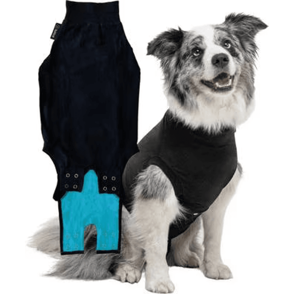
A breathable full-body recovery suit that covers a spay or abdominal incision, the vet-recommended cone alternative that lets a dog rest, walk, and go potty normally without licking or scratching the wound.
Webvet may earn a commission when you click through to Chewy, at no extra cost to you.
Dog Cyst Removal Cost
How much does cyst removal cost for a dog? For a straightforward, single cyst removed from an easy location, most owners in the U.S. pay roughly $250 to $1,000 all-in, including anesthesia. Complicated cases, difficult locations like the eyelid or face, very large cysts, or multiple cysts removed in one procedure can push the total to $1,500 or more. Regional pricing, your dog's size, and whether pre-anesthetic bloodwork is included drive most of the spread.
That removal cost range is realistic rather than best-case, and it works alongside the honest removal criteria listed in the treatment section above: you pay for surgery when a cyst ruptures, gets infected, or genuinely bothers the dog, not simply because it exists. The cost table below shows how a typical bill breaks down line by line, so you can weigh your clinic's estimate against the usual removal cost range:
| Item | Typical U.S. range | Notes |
|---|---|---|
| Exam and consultation | $50 to $250 | Higher at specialty or emergency clinics |
| Fine needle aspiration with cytology | $25 to $200 | The diagnosis step; sometimes read in-clinic for less |
| Pre-anesthetic bloodwork | $80 to $250 | Standard for seniors, recommended for all |
| Anesthesia | $90 to $300 | Scales with dog size and procedure length |
| Surgical removal, simple location | $250 to $1,000 | Single small cyst, loose skin |
| Surgical removal, complex or multiple | $1,000 to $2,500+ | Eyelid, face, paw, large or numerous cysts |
| Histopathology (biopsy of removed tissue) | $100 to $300 | Confirms the growth was benign |
| Aftercare: e-collar, meds, recheck | $30 to $150 | Often bundled |
Two ways to keep the number down without cutting corners:
- Bundle procedures. If your dog is already going under anesthesia for a dental or another surgery, ask about removing the cyst in the same session. You pay for one anesthetic episode instead of two.
- Ask early. A small cyst removed electively is cheaper than an infected, ruptured cyst removed urgently, and far cheaper than emergency-clinic pricing.
One pricing nuance owners miss: the estimate usually scales with anesthesia time, not the cyst itself. That is why a marble-sized cyst on the eyelid can cost triple what a golf-ball cyst on the flank costs, and why bundling several cysts into one procedure is dramatically cheaper than three separate surgeries.
Accident and illness pet insurance policies generally cover cyst diagnostics and removal when the cyst is not a pre-existing condition, and wellness add-ons sometimes cover part of the FNA. If a surgical estimate strains the budget, ask your clinic about payment plans or third-party financing before you delay care; a $300 elective removal beats a $1,200 emergency every time. For context on what routine visits and diagnostics run, see our guide to how much a vet visit costs.
Where Cysts Show Up on a Dog
Location changes both what a cyst is likely to be and how much it matters. This location guide covers the spots owners search most: eyelid, leg, back, ear, tail, neck, belly, and head. Each location below explains what usually grows there, which look-alikes to rule out, and whether that spot moves the cyst up the urgency list, so scan straight to the part of the body where you found your dog's bump.
Eyelid

Eyelid bumps in dogs are usually meibomian gland growths, arising from the oil glands that line the lid margin. A blocked meibomian gland forms a chalazion, a true eyelid cyst; the same glands also grow benign meibomian adenomas, which owners describe identically. An eyelid cyst matters more than its size suggests because even a small bump can rub the cornea with every blink, causing squinting, tearing, and ulcers. Never apply anything to an eyelid lump at home. These are vet-now territory, and removal, when needed, is delicate work priced at the higher end of the cost table.
Legs and Elbows
A cyst on a dog's leg is often follicular, because legs and elbows take constant pressure and friction. Elbow calluses in heavy dogs are a classic seedbed for comedones and follicular cysts. Leg cysts also get bumped and broken open more than cysts elsewhere, so they land in the "remove it" column sooner.
Back, Neck, and Shoulders
The back and neck are prime sebaceous cyst real estate: dense with oil glands and, in harness-wearing dogs, subject to daily friction. A cyst on a dog's back is usually found during petting or grooming as a pea-to-marble bump under the coat. Check that collars and harness straps are not riding directly over it.
Ears
Cysts on the ear flap or at the ear base are usually sebaceous or follicular and behave like cysts anywhere else on the body. Inside the ear canal it is a different story: cystic growths (including ceruminous gland cysts, more common in cats but seen in dogs) can narrow or block the canal, trap moisture and wax, and set up chronic ear infections that keep relapsing until the growth is addressed. Any ear bump paired with head-shaking, odor, or discharge needs prompt evaluation rather than monitoring.
Tail
The tail carries a concentration of sebaceous glands, especially at the base near the supracaudal gland. Tail cysts get whacked against furniture and door frames, so they rupture often. Recurrent tail-tip trauma can also complicate healing after removal, and enthusiastic tail-waggers may need extra bandaging or a longer restricted period to let the incision close. Ask your vet about protective options before surgery rather than after the first reopened suture line.
Belly and Stomach
Cysts on the belly sit on thin, visible skin, so owners find them early and small. The main caution: the belly is also where mammary masses appear in females, and those are never assume-benign lumps. Any bump along the mammary chains gets an FNA promptly.
Head and Face
Head and facial cysts are commonly sebaceous or follicular and are mostly a cosmetic issue until they grow near the eyes, lips, or ear openings. Facial skin is tight, so larger removals here take more surgical planning.
Which Locations Raise the Urgency
Most locations just change convenience and cost. A few change the timeline. Treat the bump as prompt-appointment territory when it sits:
- on or near the eyelid, where corneal rubbing can start within days
- in or at the entrance of the ear canal, where blockage breeds infection
- along the mammary chains in a female dog, where masses are never assumed benign
- near the anus or under the tail, where anal gland and perianal tumors mimic cysts
- anywhere it interferes with walking, chewing, vision, or elimination
Breed also stacks the deck. Cocker Spaniels, Schnauzers, Yorkshire Terriers, Shih Tzus, and Boxers are overrepresented for sebaceous and follicular cysts, and heavy breeds collect follicular cysts on calloused elbows. If you own a cyst-prone breed, expect repeat customers over the years and make the monthly lump check a permanent habit.
Wherever the bump lives, the rule does not change: one FNA turns "probably a cyst" into a diagnosis.
Cysts on Paws and Between Toes
Paw and between-the-toes swellings are their own category and usually are not true cysts at all. Most "interdigital cysts" are furuncles: deep, inflamed lesions driven by ingrown hairs, allergies, conformation, and licking, and they are treated very differently from a sebaceous cyst on the back. They tend to be red, painful, recurrent, and tied to itchy skin disease rather than a blocked gland. Because the underlying driver is usually allergic and inflammatory, the fix starts with diagnosing the itch, not cutting out the bump. We cover interdigital lesions, paw licking, and the allergy workup in our guide to itchy paws and interdigital skin problems, which is the right next read if your dog's bump is on a paw or between the toes.
Preventing Cysts and Catching Them Early
You cannot fully prevent cysts in a cyst-prone dog, but you can lower the odds and shrink the stakes:
- Groom on a schedule. Regular brushing distributes skin oils and keeps follicles clear, and it doubles as a weekly hands-on lump scan.
- Bathe appropriately, not excessively. Overbathing strips oils and can trigger rebound gland activity. Use a dog-formulated shampoo; ask your vet about follicular-flushing shampoos for comedone-prone breeds.
- Treat underlying skin disease. Allergies, seborrhea, and endocrine conditions like hypothyroidism all disrupt follicle and gland function. Managing them cuts cyst formation at the source.
- Pad the pressure points. An orthopedic bed for big dogs reduces the elbow callus trauma that seeds follicular cysts.
- Check gear fit. Collars and harnesses that rub the same spot daily create exactly the friction cysts love.
- Run a monthly lump check. Massage your dog nose-to-tail with your fingertips, note anything new in your phone with a photo and a coin for scale, and bring the log to every vet visit.
Early detection is the real win. A cyst found at pea size gets a cheap FNA and a watch-and-wait plan. The same cyst found after it ruptures gets antibiotics, surgery, and an emergency-fee line item.
Diet plays a smaller role than the internet claims, but skin health still runs on nutrition. A complete diet with adequate omega-3 fatty acids supports normal gland and follicle function, and a vet-recommended skin supplement can help dogs with chronic seborrhea. What no diet or supplement will do is shrink an existing cyst; be skeptical of any product that promises it.
The Bottom Line on Dog Cysts
A dog cyst is usually a nuisance, not a threat, and the owners who navigate them well all follow the same short playbook. Find lumps early with a monthly hands-on check. Get every new lump aspirated once, because feel is not a diagnosis. Leave cysts unsqueezed, clean up ruptures gently, and let your vet remove the ones that keep causing trouble.
The mistakes worth avoiding are just as short a list. Do not pop a cyst, ever. Do not assume a soft, movable bump is benign without an FNA. Do not spend months on home remedies while an infected or recurring cyst gets worse. And do not skip the histopathology add-on when a growth is removed; the confirmation is worth the fee.
If your dog's bump does not quite match anything in this guide, widen the lens with our lump on dog triage guide, and if the vet's needle finds something other than a cyst, our cluster covers the next steps for lipomas, mast cell tumors, and other common growths in detail.
Dog Cyst Questions Owners Ask
For a second perspective from our sister site, Petful's guide to skin cysts in dogs covers the same territory from the owner's day-to-day angle.
Frequently Asked Questions
Can a dog cyst heal on its own?
Rarely, and not in the way owners hope, because a dog cyst almost never heals fully on its own. A cyst may shrink but usually refills: it can stop growing or drain and temporarily flatten, yet the sac lining remains, so most cysts persist or come back after appearing to resolve. A small, confirmed, closed cyst can safely be monitored for the dog's lifetime. But a cyst that is infected or bothering the dog, growing, or rupturing repeatedly will not fix itself and needs veterinary treatment, usually removal.
What does a cyst look like when it comes out on a dog?
A draining cyst discharges thick material through a pore or split in the skin. Sebaceous cyst contents are grayish-white and pasty, like toothpaste or cottage cheese; follicular cyst contents are firmer and gray to yellow-brown. Blood-tinged discharge is common and not automatically alarming, but foul odor or yellow-green pus points to infection and needs a vet visit.
Are cysts painful for dogs?
An intact, uninfected cyst usually is not painful, which is why many go unnoticed until grooming. Pain shows up when a cyst becomes infected, ruptures and inflames the surrounding tissue, or sits where it is constantly pressed or rubbed. A dog that licks, chews, or flinches over a bump is telling you it has crossed from harmless to bothersome, which is exactly when vets recommend treatment.
How fast do dog cysts grow?
True cysts grow slowly, over weeks to months, and many plateau at a stable size. That slow tempo is itself diagnostic information. A lump that doubles in days to a couple of weeks, becomes ulcerated, or changes color is behaving unlike a typical cyst and should be aspirated promptly, because rapid change is one of the classic warning patterns for tumors, including mast cell tumors.

Editor
The Webvet Editorial Team is the in-house group of pet-care editors and writers behind Webvet, operated by Smart Pet Collective. The team researches, writes, and maintains Webvet's pet health, behavior, and medication content. Every article follows a defined editorial process: research from reputable veterinary and scientific sources, careful drafting, mandatory review of medical content by a credentialed veterinarian, and dated publication. Health and medication articles are medically reviewed by a licensed veterinary professional before they go live and are kept current over time.
Veterinarian · BVMS MRCVS
Dr. Pippa Elliott, BVMS, MRCVS, is a veterinarian with nearly 30 years of experience in companion animal practice. Dr. Elliott earned her Bachelor of Veterinary Medicine and Surgery from the University of Glasgow. She was also designated a Member of the Royal College of Veterinary Surgeons. Married with 2 grown-up kids, Dr. Elliott has a naughty Puggle named Poggle, 3 cats and a bearded dragon.


