Dog Eye Ulcer Pictures: What Every Stage Looks Like (Vet-Reviewed)
A vet-reviewed visual guide to dog eye ulcer pictures: what mild, deep, melting, and healing corneal ulcers look like, how the fluorescein stain works, and the red flags that make an eye ulcer a same-day emergency.
BVMS MRCVS
Medically reviewed by Dr. Pippa Elliott, BVMS MRCVS · Last reviewed

This article contains affiliate links. Webvet may earn a commission when you buy through them, at no extra cost to you.
If your dog's eye suddenly looks red, cloudy, or watery, and your dog is squinting or pawing at it, you are almost certainly looking at pain, not a passing irritation. These dog eye ulcer pictures exist for one reason: to help you recognize what a corneal ulcer looks like at each stage so you get to the vet faster, because a corneal ulcer is a same-day emergency that can deepen and blind an eye within a day or two.
- 1Photos help you gauge severity, but only a vet fluorescein stain confirms an ulcer.
- 2Superficial ulcers look like mild haze; deep ulcers show a crater or a bluish-white spot.
- 3A dark spot plugging the surface can mean perforation, a true emergency.
- 4Any painful, red, or cloudy eye needs a same-day exam.
- 5Never self-diagnose from a photo and delay care.
This is the visual guide in the WebVet dog eye ulcer cluster. It shows what mild, deep, and ruptured ulcers look like, what healing looks like in photos, and how to tell an ulcer from ordinary eye discharge. For the deeper how-and-why, we link out to the sibling guides on corneal ulcers in dogs, treatment, the day-by-day healing timeline, symptoms, ulcers that will not heal, and surgery cost.
What a Dog Eye Ulcer Looks Like (Photos)
Here is the short answer, since it is what most people came for. A dog eye ulcer usually looks like a red, cloudy, or hazy eye that the dog holds partly or fully closed, often with tearing or a thick discharge. On the clear front surface of the eye, the cornea, you may see a dull, rough, or pitted spot where the smooth shine is broken. Many dogs paw at the eye or rub it on furniture, and the eye is clearly painful.
A corneal ulcer is not a surface film or a bit of "sleep" in the corner. It is damage to the cornea itself. The VCA Animal Hospitals guide to corneal ulcers in dogs defines a true ulcer as "deeper erosion through the entire epithelium and into the stroma," meaning the injury has gone past the thin outer skin of the eye into the structural layer beneath. That is why it hurts so much and why it can get worse so fast.
The cornea is the transparent dome at the front of the eye that light passes through. When it is healthy, it is glassy and clear and you barely notice it is there. When it is ulcerated, that clarity breaks down. VCA is direct about how much this hurts and how it presents: "A corneal ulcer is very painful. Most dogs rub the affected eye with a paw or on the carpet," and, "To protect the eye, they keep the lids tightly closed. Occasionally, a discharge will collect in the corner of the eye or run down the face." VCA adds that on the eye itself, "The cornea may lose its transparency and appear cloudy, or tiny blood vessels may be seen crossing the surface." That combination of pain, a tightly held eye, discharge, and a cornea that has lost its shine is the classic ulcer picture.
The classic look of a dog eye ulcer:
- Squinting or holding the eye shut (this is pain, not sleepiness)
- Redness across the white of the eye
- Cloudiness or a bluish, hazy film over the normally clear cornea
- Excess tearing or a thick, sometimes yellow-green discharge
- A dull, rough, or dented spot on the clear surface where the shine is broken
- Light sensitivity, pawing at the eye, or rubbing the face on carpet and furniture
Not every ulcer shows all of these, and a small early ulcer can look deceptively minor. That is the trap. If your dog has any painful, red, or squinting eye, treat it as urgent and call your vet today. For a full symptom breakdown, see our companion guide on dog corneal ulcer symptoms.
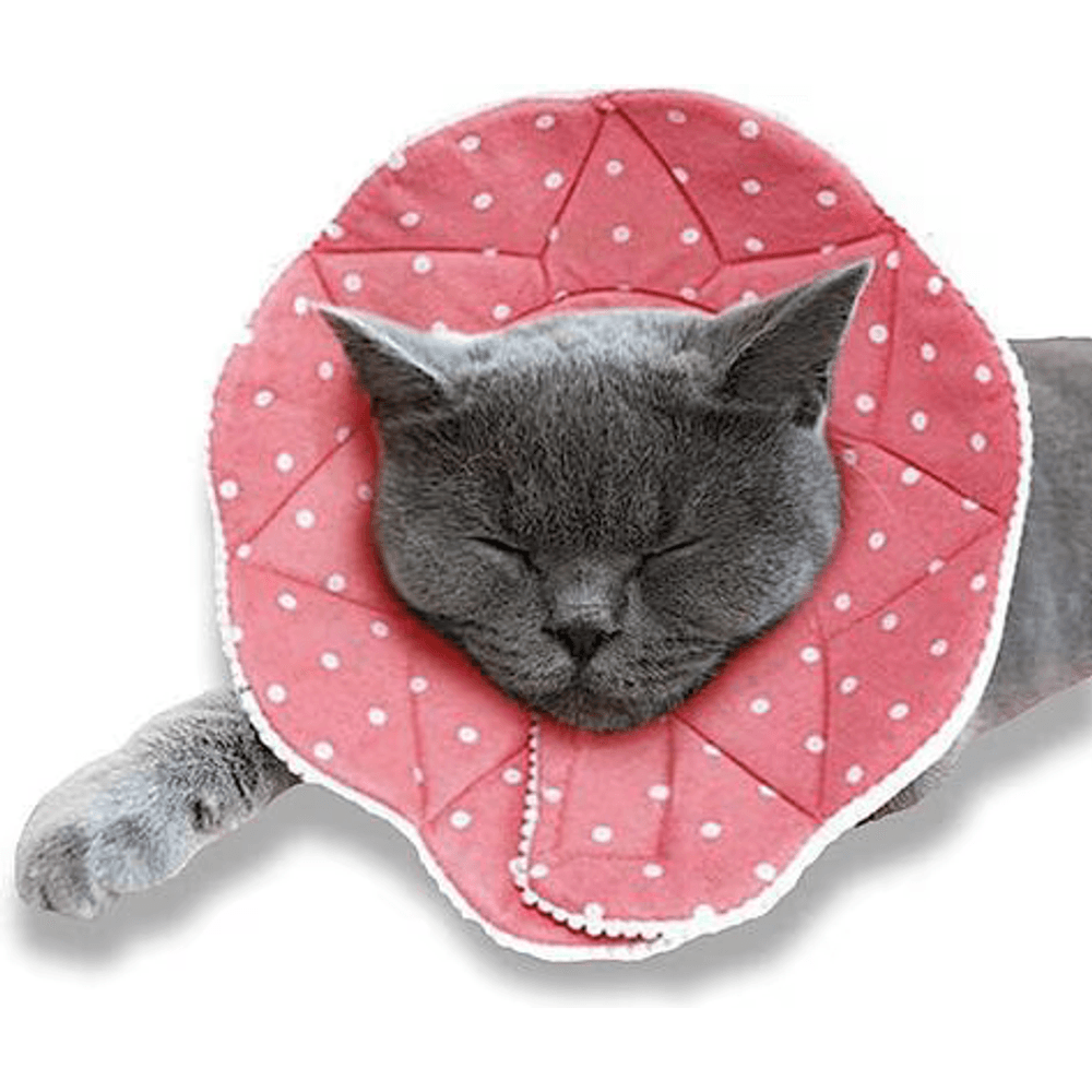
A lightweight, padded fabric cone that gently blocks a pet from pawing, scratching, or rubbing a healing eye, wound, or hot spot, and it is far softer and less stressful than a hard plastic cone. The cushioned edge and adjustable fit make it easier for dogs and cats to rest, eat, and move around while they recover.
Early-Stage vs Mild Dog Eye Ulcer: Photos and Signs

People search for early stage dog eye ulcer pictures and mild dog eye ulcer pictures because the earliest ulcers are the hardest to spot and the easiest to dismiss. That is exactly when a vet visit does the most good.
An early or mild corneal ulcer, sometimes called a superficial ulcer, involves only the outer layer of the cornea, the epithelium. In photos it tends to look like mild redness with a faint haze or dullness over part of the eye, rather than a dramatic white spot. Your dog may blink more than usual, tear a little, or squint intermittently, especially in bright light. The eye may look "off" before it looks obviously injured. This is also where mild dog eye injury pictures and true early ulcers overlap, since a fresh scratch and an early ulcer can look almost identical.
These mild ulcers are still painful and still need veterinary care, but the news at this stage is genuinely more reassuring. The Royal Veterinary College (RVC) corneal ulcer fact file notes that most ulcers heal without complications within a week when the underlying cause is gone and antibiotic drops or ointment are used. Caught early, a superficial ulcer is usually the best-case version of this problem.
What a mild or early ulcer often looks like:
- Subtle haze or dullness on one area of the cornea rather than a deep white pit
- Mild redness and a bit more tearing than normal
- Intermittent squinting or blinking, worse in bright light
- The dog is bothered but not frantic, and may still eat and play
Do not let "mild-looking" become "wait a few days." A superficial ulcer can turn into a deep or infected one quickly, and only a fluorescein stain tells your vet how deep it really is. A mild-looking eye is still a same-day call. The point of catching it early is that early ulcers are far easier to heal than the deep ones in the next section.
Deep, Melting, and Ruptured Ulcers: What Severe Looks Like


This is the part that shows why "wait and see" is dangerous. When people search deep corneal ulcer dog, ruptured corneal ulcer dog, or descemetocele dog, they are usually looking at an eye that has crossed from painful into vision-threatening.
A deep or stromal ulcer has eroded down into the structural stroma of the cornea. In photos it looks like a visible crater or pit in the surface, often with pronounced white or gray cloudiness around it and heavy discharge. The eye may look sunken at the ulcer site. VCA describes this progression through the layers of the cornea: "If the erosion goes through the epithelium and stroma to the deepest level of Descemet's membrane, a descemetocele is formed," which it calls "a serious condition." Descemet's membrane is the last thin layer before the inside of the eye, so once an ulcer reaches it the eye is close to giving way.
A descemetocele is the near-perforation stage: the ulcer has eaten through everything except that final Descemet's membrane, which bulges forward like a clear blister at the bottom of the crater. It is one step away from rupture. The Merck Veterinary Manual section on ophthalmic emergencies explains that deep ulcers, descemetoceles, and ulcers that have already ruptured with iris prolapse require immediate surgical support of the fragile globe because they seriously compromise the integrity of the eye.
A melting ulcer is a specific, fast-moving emergency where enzymes break the cornea down almost in front of your eyes. In photos it looks gray, gelatinous, and gooey, as if the surface of the eye is dissolving or sliding. Melting ulcers can go from bad to ruptured in hours, not days. These are the dog eye ulcer rupture symptoms owners fear, and they are a reason to go straight to an emergency hospital.
A ruptured (perforated) ulcer is the worst case. The cornea has broken open, fluid leaks out, and part of the iris may plug the hole and appear as a dark spot or bump on the surface. The eye may look collapsed or distorted. VCA is blunt about the stakes: "If Descemet's membrane ruptures, the liquid inside the eyeball leaks out, the eye collapses, and irreparable damage occurs." A ruptured globe is what "losing the eye" actually looks like, and it is why Merck lists ruptured ulcers with iris prolapse among the emergencies that need immediate surgery.
Severe ulcer red flags in photos and in person:
- A visible crater, pit, or divot in the clear surface of the eye
- Dense white, gray, or blue cloudiness, sometimes with a gooey or melting look
- A clear blister-like bulge at the base of a crater (a possible descemetocele)
- A dark spot or bump on the surface, or an eye that looks collapsed or misshapen (possible rupture with iris prolapse)
- Severe pain, the eye held tightly shut, heavy or bloody discharge
If your dog's eye looks anything like this, this is an emergency now, not tomorrow. Go to your veterinarian or the nearest emergency animal hospital. The surgery your dog may need, and what it costs, is covered in a dedicated sibling guide.
Presoaked sterile pads that gently wipe away everyday debris, discharge, and tear stains from around a dog's or cat's eyes as part of routine grooming. An easy way to keep the eye area clean and comfortable between baths. For routine cleaning only, not for treating an injured or infected eye, which needs a vet.
Dog Eye Ulcer Healing Stages (Pictures)

Signs a dog eye ulcer is healing and dog eye ulcer healing stages images are among the most-searched parts of this topic, because once you are past the scary part you want proof it is getting better. This page shows you what healing looks like. For the full day-by-day timeline and prognosis, the dog eye ulcer healing stages guide owns the detailed schedule; here we focus on the pictures.
The short version of the timeline: a simple superficial ulcer usually re-surfaces fast. VCA notes that corneal abrasions and superficial ulcers generally heal within three to five days, and the RVC fact file says most ulcers heal within about a week when treated and the cause is removed. Deep, infected, indolent, or melting ulcers heal much more slowly and may need surgery, so a longer dog eye ulcer healing time is not automatically bad news but it does mean staying in close contact with your vet.
Here is what improvement tends to look like as the days pass.
What a healing ulcer looks like in photos
| Stage | What you often see in photos | What it means |
|---|---|---|
| Active ulcer (day 0) | Red, cloudy eye, held shut, a dull or pitted spot, discharge | The ulcer is open and painful; treatment just starting |
| Early healing (first days) | Less squinting, less discharge, eye opening more, a little redness | The surface is starting to re-cover; pain easing |
| Vascularization | Tiny red blood vessels creeping in from the edge toward the ulcer | The body is delivering healing cells; a normal, good sign |
| Re-surfacing | Cloudiness shrinking, the clear shine returning to more of the cornea | New epithelium is closing the defect |
| Faint scar / haze | A small gray haze or faint scar where the ulcer was | Often the final, harmless footprint of a healed ulcer |
Positive signs your dog's ulcer is healing:
- Your dog opens the eye more and squints less
- Less tearing and discharge than a few days ago
- The eye looks less red overall
- Cloudiness is shrinking and more of the cornea looks clear
- Your dog seems more comfortable, less pawing and rubbing
Warning signs it is NOT healing and you should call the vet again promptly:
- Increasing pain, redness, squinting, or discharge
- The cloudy or pitted area getting bigger or deeper
- A gooey, melting, or bulging look developing
- No improvement after the number of days your vet expected
Small blood vessels growing across the cornea can look alarming in photos, but they are usually your dog's body doing exactly the right thing. Regrowth of clear surface and a fading of the haze are the goal. If things look worse instead of better, that is a same-day call. Some ulcers stall for reasons we cover under ulcers that will not heal.

How Vets Diagnose an Eye Ulcer: The Fluorescein Stain (Green Glow)

Those striking fluorescein stain dog eye photos, where a spot on the eye glows bright green, are not a special effect. That green glow is the single most important test for diagnosing a corneal ulcer, and it is the reason a vet can confirm in seconds what you cannot confirm at home.
Here is how it works. VCA describes the process plainly: "A drop of this orange-colored stain is placed on the cornea. The dye will adhere to ulcerated areas and turn green." The healthy, intact surface of the cornea repels the dye, so it washes off clear. But wherever the protective outer layer is broken, the exposed tissue underneath grabs the stain and lights up green, often under a blue light. The size and shape of the green area shows the vet exactly where the ulcer is and how big it is.
Depth matters as much as size. The Merck Veterinary Manual notes that ulcer depth must be assessed with magnification and a slit lamp, and that a Seidel test, done with the same fluorescein stain, checks for active fluid leakage if the vet suspects the cornea has actually ruptured. This is why an eye that "looks okay" to you still needs a professional exam: a small green spot and a deep, sight-threatening crater can look similar from across the room but are worlds apart in urgency.
Why you cannot do this at home:
- The stain and blue light, and the trained eye to read them, are the diagnostic tools, not a phone camera
- Depth, infection, and melting are judged with equipment you do not have
- Guessing wrong, especially reaching for the wrong drops, can cost your dog the eye
The takeaway is simple. Pictures like the ones in this article are for recognition, not diagnosis. Only a fluorescein stain performed by your veterinarian confirms an ulcer and tells you how serious it is.

Presoaked wipes that gently clean the fur and skin around a dog's or cat's eyes, lifting away tear stains, discharge, and daily debris as part of routine grooming. A quick, no-rinse way to keep the eye area clean and tidy between baths. For routine cleaning only, not for treating an injured or infected eye, which needs a vet.
When a Dog Eye Ulcer Is an Emergency
If you take one thing from this whole guide, take this: a painful, red, squinting, cloudy, or heavily tearing eye is an emergency, and the safest assumption is always "vet today." A dog eye ulcer emergency is defined by how fast the eye can deteriorate, not by how bad it looks in the first hour.
Corneal ulcers are dangerous because of how fast they can change. A superficial ulcer can become a deep, infected, or melting ulcer within a day or two, and the Merck Veterinary Manual is clear that descemetoceles and ruptured ulcers with iris prolapse "require immediate surgical support of the fragile globe." VCA confirms the ceiling on the risk: once Descemet's membrane ruptures, "the liquid inside the eyeball leaks out, the eye collapses, and irreparable damage occurs." Merck also notes that deep central ulcers "can markedly impair vision" even short of that. This is not a problem that rewards patience.
Go to a vet or emergency hospital right away if you see:
- A crater, pit, or divot in the surface of the eye
- A gooey, melting, or dissolving look to the cornea
- A bulging blister at the base of an ulcer, or a dark bump on the surface
- The eye looking collapsed, distorted, or larger or smaller than the other
- Severe pain, an eye clamped shut, or bloody discharge
- Sudden vision loss or bumping into things
What slows down healing (and what to avoid)
People search what slows down dog eye ulcer healing and "dog eye ulcer won't heal" because a stalled ulcer is frightening, and a corneal ulcer dog not healing on schedule is a real red flag. Healing is delayed by ongoing causes that have not been fixed (an inturned eyelid, an extra lash, a trapped foreign body like a thorn), by infection, by dry eye, and by indolent (SCCED) ulcers that will not stick down on their own. The RVC fact file lists these exact culprits and notes that some, such as melting ulcers, eyelid abnormalities, retained foreign bodies, and SCCEDs, often need surgery rather than drops alone. Central ulcers can also be slower, because, as Merck notes, they need more time and blood-vessel in-growth to heal.
The single most important thing owners can do to avoid slowing healing: never put anything in the eye that your vet did not prescribe for this ulcer. That means:
- No steroid-containing eye drops. Many combination eye or ear medications left over from an old problem contain a steroid, and a steroid on an active ulcer can make it worse and even melt or perforate. When in doubt, do not use it.
- No human eye drops or redness relievers.
- No leftover medications from a previous pet or a previous problem.
- Do not rub, wipe, or flush the eye with anything except plain saline, and only if your vet tells you to.
If a stalled ulcer keeps you up at night, our sibling guide on a corneal ulcer that is not healing walks through indolent and refractory ulcers in depth.

A sterile lubricating gel that soothes and moisturizes dry, irritated eyes and helps support the tear film in dogs and cats prone to dryness. A gentle, vet-shelf staple for everyday eye comfort. It is not a treatment for an eye injury or infection, so a painful, red, or cloudy eye still needs a same-day vet visit.
What Causes Eye Ulcers in Dogs (and At-Risk Breeds)

Understanding what causes dog eye ulcer helps you spot risk before an injury and know why some dogs are back at the vet again and again. The single biggest cause is trauma. The RVC fact file states that most ulcers in dogs and cats are the result of trauma, such as scratches during walks, thorns, playing, or scratches from other animals. VCA adds blunt trauma from eye rubbing, chemical burns from substances like shampoo or drywall dust, and lacerations from sharp objects.
Beyond direct injury, common dog eye ulcer causes include:
- Dry eye (keratoconjunctivitis sicca), where too little tear film leaves the cornea exposed and prone to ulceration
- Eyelid and eyelash abnormalities, such as entropion (an inturned lid) or ectopic cilia (lashes growing in the wrong place) that scrape the cornea
- Foreign bodies, like a grass seed or thorn trapped behind the lid
- Infection, bacterial or viral, which can start or worsen an ulcer
- Chemicals and irritants splashed into the eye
Breeds at higher risk
Owners searching eye ulcer boxer dog or shih tzu dog eye ulcer are usually right to be worried, because flat-faced (brachycephalic) breeds are genuinely more prone to corneal ulcers. VCA specifically notes that brachycephalic breeds such as pugs and bulldogs are more susceptible because of their anatomy: prominent, exposed eyes, shallow sockets, and often an incomplete blink that leaves the cornea drying and undefended. Merck adds that brachycephalic dogs and dogs with dry eye are especially vulnerable to stromal loss from infectious keratitis.
Higher-risk dogs include:
| Group | Examples | Why they are at risk |
|---|---|---|
| Brachycephalic (flat-faced) | Boxer, Pug, Shih Tzu, Bulldog, Pekingese, Boston Terrier | Large, prominent eyes and incomplete blink leave the cornea exposed and dry |
| Dogs with eyelid conformation issues | Many brachycephalics, plus some larger breeds | Entropion or extra lashes rub the cornea constantly |
| Dogs prone to dry eye | Cocker Spaniel, Bulldog, West Highland White Terrier, others | Low tear film leaves the cornea unprotected |
| Older dogs | Any breed | Indolent (SCCED) ulcers become more common with age |
Boxers in particular are the classic example of indolent ulcers, superficial ulcers that will not heal normally, which is why the breed comes up so often in searches. If you own a flat-faced or dry-eye-prone dog, treat any red or squinting eye as urgent, because these dogs have the least margin for a wait-and-see approach.
Treatment and the 1-2-3 Rule (Why You Must See a Vet)

A quick note on scope: this page shows you what an ulcer looks like. The full dog eye ulcer treatment protocol lives in our dog eye ulcer treatment guide, and cost details are in the surgery cost guide. Here is the essential summary and the answer to the popular 1-2-3 rule question, framed as what your vet will do, never as a DIY plan.
How do you treat an ulcer on a dog's eye? In veterinary hands, a simple superficial ulcer is usually treated with antibiotic eye drops or ointment to prevent infection, plus pain relief, while the cornea re-surfaces on its own. The RVC fact file confirms that most ulcers heal with antibiotic drops or ointment as long as the underlying cause is gone. Deeper, infected, indolent, or melting ulcers may need extra medications, a procedure to help an indolent ulcer heal, or surgery such as a conjunctival graft. Merck notes that ulcers with at least 50% stromal loss should be assessed by an ophthalmologist for surgical repair.
You may also see a soft contact lens (bandage lens) or an E-collar (cone) used to protect the eye. The cone is not optional cruelty; it stops your dog from rubbing the ulcer open again and is one of the most important parts of home care your vet will insist on.
What is the 1-2-3 rule for corneal ulcers?
The 1-2-3 rule is a veterinary rule of thumb, not a home treatment. Broadly, it is used by some vets and eye specialists as a quick guide to categorize a corneal ulcer by how many days it has been present, how deep it is, and how many follow-up rechecks or escalating steps it needs, so that an ulcer not improving on schedule gets escalated (for example, referred to an ophthalmologist or considered for surgery) rather than left on the same drops indefinitely. Different clinics phrase it slightly differently. The practical point for you as an owner is simple: ulcers are tracked on a timeline, and any ulcer that is not clearly improving on schedule needs to be re-checked and escalated by a vet, not managed at home.
What you should never do at home:
- Never use steroid-containing drops, human eye drops, or leftover medications
- Never try to flush, wipe, or medicate the eye without your vet's specific instructions
- Never skip the cone or the rechecks your vet schedules
Everything on the treatment side starts with a veterinary exam and a fluorescein stain. If your dog has any painful or cloudy eye, the single best treatment you can provide is a same-day trip to the vet.

Dog Eye Ulcer vs Eye Discharge: How to Tell the Difference

One of the most useful things a picture guide can do is help you separate a true ulcer from a simpler eye problem like plain watery discharge or conjunctivitis. They can look similar at a glance, but an ulcer is far more urgent.
| Feature | Corneal ulcer | Plain discharge or mild conjunctivitis |
|---|---|---|
| Pain | Marked; squinting, holding eye shut, pawing | Usually mild or none |
| The clear cornea | Cloudy, hazy, or with a visible dull or pitted spot | Usually stays clear and shiny |
| Redness | Often significant | Can be mild to moderate |
| Discharge | Tearing and often thick discharge | Watery to mucousy discharge |
| Urgency | Same-day emergency | Still worth a vet call, less time-critical |
| Confirmed by | Fluorescein stain (green glow) at the vet | Exam; often no staining uptake |
The honest bottom line: you cannot reliably tell the difference at home, and you should not try to. When there is pain, cloudiness, or squinting, assume the more serious problem and see your vet the same day. This differentiation is about knowing how urgent to be, not about deciding to stay home.
The Bottom Line on Dog Eye Ulcer Pictures
Dog eye ulcer pictures are a recognition tool, full stop. A red, cloudy, squinting, or heavily tearing eye means your dog is in pain and needs a veterinarian today, because a corneal ulcer can deepen and perforate within 24 to 48 hours and threaten the eye itself. Mild ulcers usually heal within about a week with prompt care; deep, melting, or ruptured ulcers can cost the eye. Only a fluorescein stain at the vet confirms an ulcer, and the most dangerous thing you can do at home is reach for the wrong drops. When in doubt, go in.
For the deeper dives, use the sibling guides linked throughout: the corneal ulcer overview, symptoms, healing stages timeline, treatment, ulcers that will not heal, and surgery cost.
Frequently Asked Questions
What does an ulcer on a dog's eye look like?
An ulcer on a dog's eye usually looks like a red, cloudy, or hazy eye held partly or fully shut, with tearing or thick discharge, and often a dull, rough, or pitted spot on the normally clear front surface of the eye. VCA describes an ulcer as erosion through the entire epithelium and into the stroma, notes it is very painful so the dog keeps the lids tightly closed, and says the cornea may lose its transparency and appear cloudy while discharge collects at the corner of the eye. A mild early ulcer may just look like faint haze and mild redness, while a deep one shows a visible crater. Any of these means a same-day vet visit.
Will an eye ulcer in a dog heal on its own?
Not safely, and not without a vet. A simple, superficial ulcer often heals quickly once treated: VCA notes superficial ulcers generally heal in three to five days, and the RVC says most heal within about a week with antibiotic drops and the cause removed. But you cannot tell at home whether an ulcer is superficial or deep, and an untreated ulcer can become infected, melt, or perforate. The correct answer is to have a vet confirm it with a fluorescein stain and prescribe treatment, not to hope it clears up on its own.
How do you treat an ulcer on a dog's eye?
Treatment is prescribed by a vet after a fluorescein stain. A superficial ulcer is typically treated with antibiotic eye drops or ointment plus pain relief while it re-surfaces, and the RVC confirms most ulcers heal this way when the cause is gone. Deep, infected, indolent, or melting ulcers may need extra medication, a procedure, or surgery such as a conjunctival graft; Merck notes ulcers with 50% or more stromal loss should be assessed for surgery. A cone and rechecks are part of care. Never use leftover, human, or steroid drops at home.
What slows down dog eye ulcer healing?
Healing stalls when an underlying cause has not been fixed or when the ulcer is a difficult type. The RVC fact file points to eyelid abnormalities (like entropion), retained foreign bodies (a thorn), dry eye, infection, melting ulcers, and indolent SCCED ulcers, several of which need surgery rather than drops alone. Merck adds that central ulcers heal more slowly because they need more time and blood-vessel in-growth. The other major cause of delayed healing is owners using the wrong products, especially steroid drops, which can make an ulcer worse.
Can a dog's eye ulcer heal itself?
A shallow, superficial ulcer can re-surface on its own within days once the cause is removed, but it still needs a vet, because only a stain shows how deep it is and because pain and infection need managing. Deep, melting, indolent, or infected ulcers will not heal on their own and can worsen fast: VCA warns that if the ulcer erodes to Descemet's membrane and it ruptures, the eye collapses and irreparable damage occurs, and Merck lists ruptured ulcers with iris prolapse among the emergencies needing immediate surgery. So while some ulcers technically re-surface without surgery, healing itself should never mean skipping the vet.
What is the 1-2-3 rule for corneal ulcers?
The 1-2-3 rule is a veterinary shorthand, not a home remedy. It is used loosely to remind clinicians to track a corneal ulcer on a timeline by how long it has been present, how deep it is, and how many rechecks or escalating steps it needs, so any ulcer not improving on schedule gets escalated (for example, referred to an ophthalmologist or considered for surgery) instead of being left on the same drops. Wording varies by clinic. For owners, the practical meaning is that ulcers are followed on a schedule, and any that is not clearly improving needs a professional re-check, not more waiting.
How do I help my dog's eye ulcer?
The most helpful thing you can do is get to the vet the same day and then follow their plan exactly. At home that usually means giving the prescribed drops or ointment on schedule, keeping the cone on at all times so your dog cannot rub the eye, restricting rough play, and going to every recheck. Watch for worsening pain, redness, cloudiness, or a melting or bulging look, and call immediately if you see them. Just as important is what not to do: never use leftover, human, or steroid eye drops, and do not wipe, rub, or flush the eye unless your vet specifically tells you to.

Editor
The Webvet Editorial Team is the in-house group of pet-care editors and writers behind Webvet, operated by Smart Pet Collective. The team researches, writes, and maintains Webvet's pet health, behavior, and medication content. Every article follows a defined editorial process: research from reputable veterinary and scientific sources, careful drafting, mandatory review of medical content by a credentialed veterinarian, and dated publication. Health and medication articles are medically reviewed by a licensed veterinary professional before they go live and are kept current over time.

Veterinarian · BVMS MRCVS
Dr. Pippa Elliott, BVMS, MRCVS, is a veterinarian with nearly 30 years of experience in companion animal practice. Dr. Elliott earned her Bachelor of Veterinary Medicine and Surgery from the University of Glasgow. She was also designated a Member of the Royal College of Veterinary Surgeons. Married with 2 grown-up kids, Dr. Elliott has a naughty Puggle named Poggle, 3 cats and a bearded dragon.
Related reading

Dog Eye Ulcer Surgery Cost: 2026 Price Guide by Procedure

Dog Eye Ulcer Won't Heal? Causes, Danger Signs, and How Vets Fix It

Signs a Dog Eye Ulcer Is Healing: Stages, Timeline, and Red Flags
