Salivary Mucocele in Dogs: Symptoms, Surgery, and Recovery
A jaw or neck swelling needs a diagnosis. Learn how vets identify a salivary mucocele, why drainage can fail, and what surgery, costs and recovery may involve.

BVMS MRCVS
Medically reviewed by Dr. Pippa Elliott, BVMS MRCVS · Last reviewed

This article contains affiliate links. Webvet may earn a commission when you buy through them, at no extra cost to you.
A salivary mucocele in dogs is a pocket of leaking saliva beneath the skin or inside the mouth or throat. It often appears as a soft swelling near the jaw or neck. A dog may otherwise seem comfortable, but swelling that interferes with swallowing or breathing needs urgent veterinary attention.
The important decisions are whether the airway is affected, what the swelling contains, and which gland or duct is leaking. A photograph or a lump that feels soft cannot settle those questions. Treatment usually addresses the source of the leak, rather than repeatedly emptying the pocket.
 Sponsored
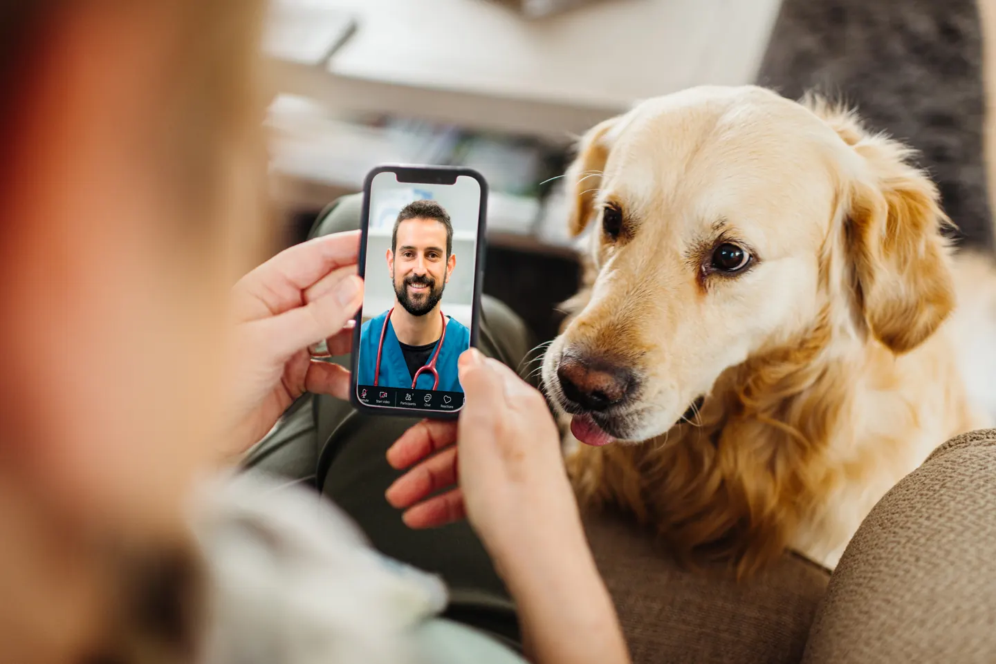
Have a health question about your dog?
Talk to a licensed vet at Dutch over video or message, including nights and weekends.
Use code Pets30
Plans start at $11/month · covers up to 5 pets
- Licensed vets in most states
- Rx for 150+ conditions
- 4.7 on Trustpilot
What a salivary mucocele is
Leaking saliva causes inflammation and a pocket of fluid. Arrange veterinary assessment for a new jaw or neck swelling; difficulty breathing is an emergency. Do not squeeze, puncture or try to drain the lump yourself.
Salivary glands make saliva and send it into the mouth through ducts. When that system is damaged, saliva can escape into nearby tissues. The body develops a wall of inflammatory tissue around the collection. You may hear the words sialocele, salivary mucocele or salivary cyst used for the same problem, although it is not a true gland-lined cyst.
Several paired glands contribute saliva. The mandibular and sublingual glands are commonly involved in these cases; parotid and zygomatic glands occupy different positions. Knowing which side first enlarged can help the veterinarian, particularly after swelling spreads toward the middle of the neck.
For a planned visit with a small dog, a correctly sized Frisco Travel Safety Dog & Cat Carrier can provide secure transport. Check that the dog fits comfortably and that the journey follows the clinic's advice. A medium carrier is not suitable for every dog, especially a large patient.
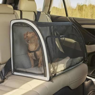
Secure transport for a small dog that fits the linked medium carrier.
Webvet may earn a commission when you click through to Chewy, at no extra cost to you.
What to tell the clinic
Record when the swelling appeared, its original side, changes in size and any chewing injury or neck trauma. Mention drooling, difficulty eating, a changed bark or noisy breathing. Photographs taken without handling the lump can document change, but they do not replace an examination. The broader guide to a lump on a dog explains why different masses may need different tests.
Neck swelling and urgent swallowing or breathing signs
Swollen salivary glands in dogs, including a puppy, need veterinary examination because infection, obstruction and other masses can resemble a mucocele. A sialocele, often called a salivary cyst, typically produces a gradually enlarging, soft neck swelling; symptoms depend on its location. It may be painless, so normal play or appetite does not prove that it is harmless. A ranula is a saliva collection beneath or beside the tongue; pharyngeal involvement affects the throat and can be much harder to see from outside.
Once the clinic has assessed urgency, a comfortable resting area helps you observe eating and breathing without repeatedly handling the neck. For a large dog, the FurHaven NAP Ultra Plush Full Support Deluxe bed in Jumbo is a floor-level option. Check its dimensions and cover-care directions; a bed does not relieve pressure from a growing swelling.
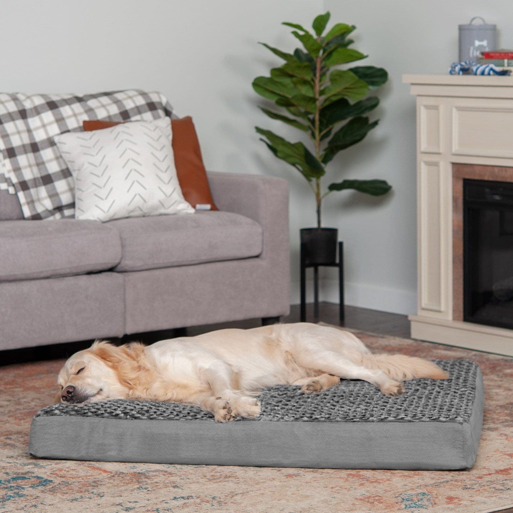
A floor-level resting option for a large dog; check the Jumbo dimensions and cover care.
Webvet may earn a commission when you click through to Chewy, at no extra cost to you.

Figure 1 from Kazemi, Doustar and Assadnassab's published case (2012), reproduced under the paper's Creative Commons Attribution terms. This German Shepherd had chronic neck swelling and is undergoing veterinary aspiration. The image is a documented example, not a way to diagnose another dog's lump.
| Swelling location and next step | What an owner may notice | Appropriate action |
|---|---|---|
| Cervical: upper neck or under the jaw | A new or enlarging lump, sometimes without obvious illness | Arrange an examination; report its original side and changes |
| Sublingual: floor of the mouth | Trouble chewing, swallowing difficulty, drooling or blood after trauma to the swelling | Contact the clinic promptly; do not force the mouth open or puncture it |
| Pharyngeal: throat | Swallowing difficulty, changed breathing or respiratory distress, sometimes without an external lump | Breathing difficulty requires emergency veterinary care |
| Zygomatic: near the cheek and eye | Swelling below the eye or facial asymmetry | Seek examination; dental, eye and other facial disorders can look similar |
Can a dog live with an untreated mucocele?
A dog may live with a cervical swelling for a time, but that is not a safe prediction for an individual case. If a mucocele is left untreated, continuing leakage can enlarge the pocket. Throat involvement can obstruct the airway and become fatal. There is no dependable number of hours, days or months before an untreated mucocele becomes dangerous; location and current function matter more than a calendar.
Struggling for breath, collapse or inability to swallow warrants immediate veterinary help. Do not offer food, water or oral medicine to a dog struggling to swallow. Call ahead so the team can prepare. ACVS guidance specifically identifies pharyngeal mucoceles as a potentially life-threatening airway problem.
How a veterinarian diagnoses the swelling
The examination considers the mouth, teeth, salivary glands, lymph nodes and surrounding tissues. Owners cannot reliably distinguish a swollen salivary gland from a lymph node by location or feel. The normal gland's size also does not provide a useful home threshold for deciding whether a lump needs testing.
Veterinary aspiration removes a small fluid sample with sterile equipment. The clinician assesses the material and may examine cells under a microscope, a test called cytology. Saliva-like fluid supports a mucocele diagnosis; findings suggesting infection, an abscess or a tumor change the investigation. A blood test alone does not establish what a neck mass contains.
When imaging or additional testing helps
Ultrasound or CT may help define an unusual, deep or recurrent swelling and its relationship to important structures. Imaging is not automatically necessary for every straightforward case. Sedation or anesthesia may be needed to examine a hidden throat swelling safely, especially when opening the mouth causes distress.
Other salivary disorders include inflammation, infection, duct stones and tumors. Salivary gland hyperplasia means increased gland tissue, not automatically a saliva pocket. A draining tract or fistula likewise needs examination rather than repeated wiping and observation. These possibilities explain why an “overactive gland” is not a sufficient diagnosis for drooling or swelling.
For example, a published series of three unusual mucoceles documented mineralized walls and more complex investigations. Such cases illustrate exceptions; they do not mean that every soft swelling will harden. Routine saliva-amylase questions or internet gland diagrams cannot replace sampling and interpretation of the actual lesion.
 Sponsored
Vet care that's always there
Unlimited video calls and messaging with licensed vets for less than a typical vet visit. One membership covers up to five pets.
Use code Pets30
Plans start at $11/month · covers up to 5 pets
- Licensed vets in most states
- Rx for 150+ conditions
- 4.7 on Trustpilot
Treatment, drainage, and gland-removal surgery
The usual definitive treatment is removal of the affected gland and associated duct tissue, called sialoadenectomy. The surgeon selects the approach after identifying the likely source and extent of the leak. Mandibular and sublingual glands are often removed together because of their close anatomical relationship.
This is delicate neck surgery involving nearby nerves and vessels, so referral to a surgical specialist may be appropriate. Removing a diseased gland does not usually leave a dog unable to make enough saliva. Other glands continue to function, and a normal life is possible after successful treatment.
If tablets are prescribed, ask the pharmacy whether they can safely be taken out of their original packaging. Where storage permits, an Ezy Dose Weekly 7-Day Dog Pill Organizer can help track a clear schedule. Keep the prescription label and instructions, and keep all medication out of reach of children and animals.
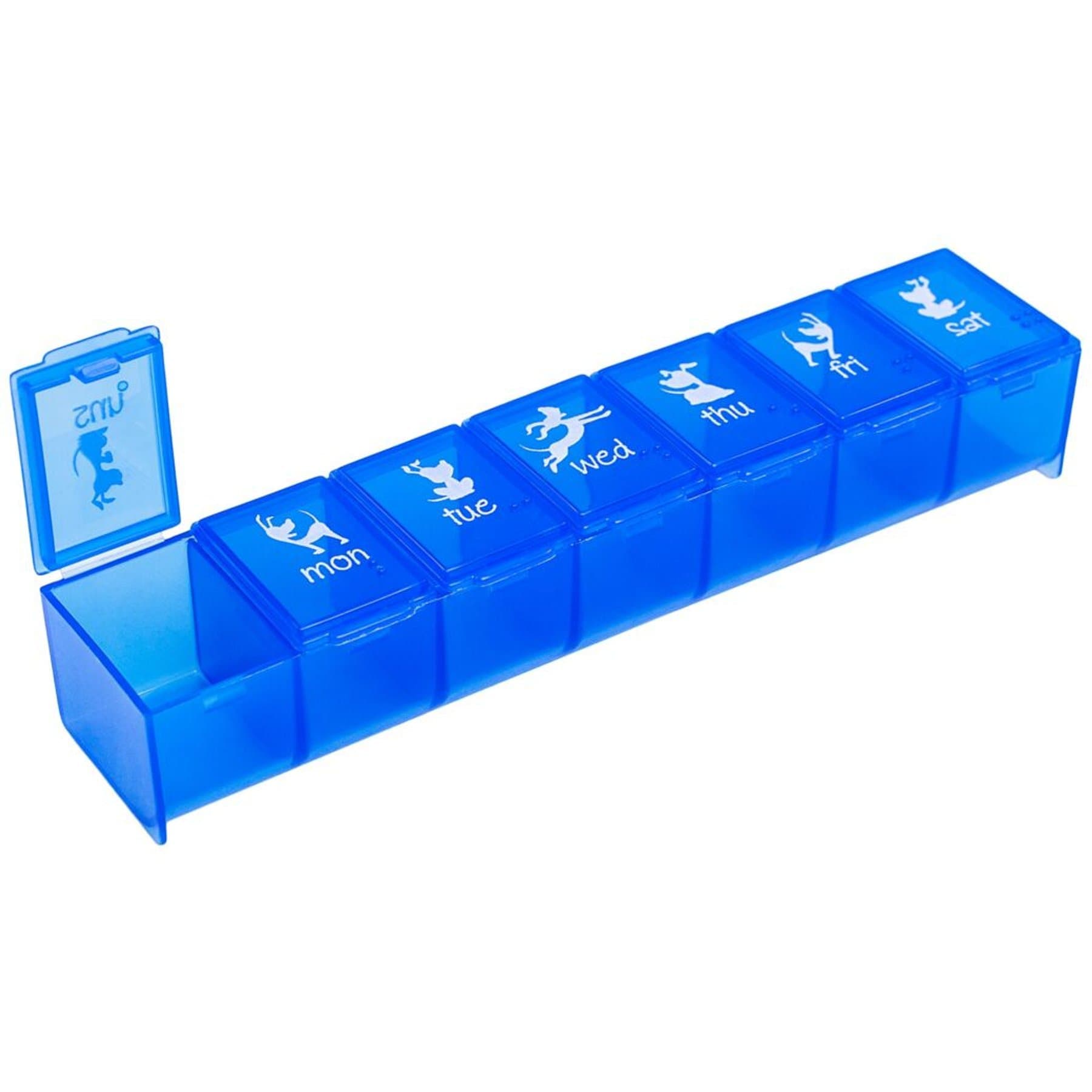
Organize prescribed tablets only when the pharmacy confirms suitable storage.
Webvet may earn a commission when you click through to Chewy, at no extra cost to you.
| Intervention | Main purpose | Important limit |
|---|---|---|
| Diagnostic aspiration | Sample the swelling to investigate its contents | Sampling does not repair the leak |
| Temporary drainage | Reduce the collected fluid | Fluid may return; repeat procedures introduce infection risk |
| Gland removal | Address the source of leaking saliva | Requires an individualized surgical and anesthetic plan |
| Antibiotics | Treat infection when indicated | Do not reliably seal a damaged salivary duct |
Why drainage is not the same as fixing the leak
Diagnostic aspiration and temporary drainage have different purposes. Aspiration obtains information; drainage may reduce a fluid pocket for a time. Neither necessarily stops the damaged gland or duct from supplying more saliva. Repeated drainage can allow recurrence and introduce infection, making subsequent care more difficult.
A ranula may also need a surgically created opening into the mouth, called marsupialization. Do not pop an oral mucocele. That is different from a controlled veterinary procedure and risks injury and infection. A blocked duct, ruptured mucocele or spontaneously draining lump still needs a veterinary plan; an opening in the skin does not prove that the underlying problem has healed.
Antibiotics, natural remedies and waiting for it to disappear
Antibiotics may be needed for a documented or suspected infection, but they do not reliably repair a leaking duct. An uncomplicated saliva pocket is not automatically infected. Do not reuse a previous antibiotic or assume that swelling shrinking during treatment proves a permanent cure.
There is no established home remedy that safely removes a mucocele or reliably seals the leak. Massage, puncturing, squeezing, applying caustic substances or attempting oral drainage can injure sensitive tissue and delay care. A swelling that temporarily gets smaller still needs the recommended reassessment rather than an assumption that it has healed on its own.
Occasionally a veterinarian may consider a conservative approach when surgery is unsuitable or being planned. Discuss its goal, recurrence risk and reassessment threshold. A report of a refractory recurrent sialocele describes highly individualized specialist care; it is not a home-treatment recipe or evidence that those interventions should replace standard surgery.
Surgery cost and questions for an itemized estimate
Salivary mucocele surgery cost depends on the hospital, location, emergency needs, imaging, anesthetic assessment and complexity of the gland removal. A general-practice quote and a specialist referral quote may include very different services. A single national average would obscure those differences.
For feeding after discharge, ask what texture and portion size the surgeon allows. A small OurPets Durapet stainless bowl, 0.75 cups can hold measured small-dog portions and is easy to wash. It is not a full-size meal bowl for a large dog, and changing bowls will not correct swallowing difficulty.
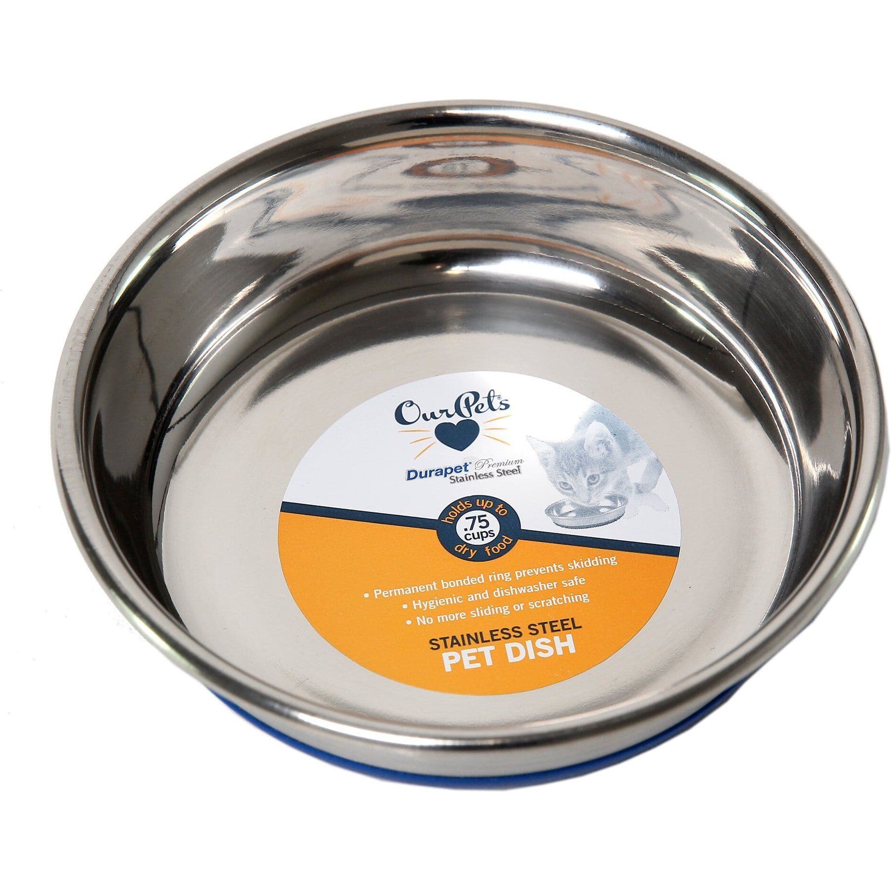
A small washable bowl for measured portions after the surgeon permits oral feeding.
Webvet may earn a commission when you click through to Chewy, at no extra cost to you.
Ask for an estimate showing:
- Consultation, sampling and any additional imaging.
- Preanesthetic tests, anesthesia and surgical fees.
- Hospitalization, pain relief and other prescribed medicines.
- Tissue analysis, drain removal and scheduled rechecks.
- What happens financially if the findings or recovery require additional care.
Before the operation, identify who will answer recovery questions outside normal hours. Ask whether an unexpected swelling recheck is included and whether a second procedure, if needed, would require a new estimate. Keep written discharge and cost documents together so the next clinician can see what was performed, not just the original diagnosis.
Comparing quotes in different countries
A price from the UK, Canada, Australia, India or the Philippines should retain its currency, date and inclusions. Converting it into dollars does not make it a comparable US quote. Ask nearby clinics whether the consultation and specialist charges are separate, and whether the estimate covers the same proposed operation and follow-up.
Tell the team early if cost is a constraint. That allows a realistic discussion of referral options, payment arrangements or available assistance while the dog is being assessed. Affordability changes planning; it does not make airway obstruction safe to watch at home.
Recovery, incision care, and recurrence
Recovery from salivary gland removal involves both the incision healing and the dog returning to comfortable eating and activity. Ask the surgeon for the expected milestones for this particular operation, including rechecks and removal of sutures or a drain. An internet recovery date cannot account for an infected, recurrent or unusually extensive mucocele.

Follow the individual discharge instructions for:
- Food texture, portions and when oral feeding can resume.
- Each medicine, its purpose and what to do about a missed dose.
- Exercise restrictions and prevention of scratching or rubbing.
- Incision observations and any prescribed drain or bandage care.
- The recheck appointment and signs that require an earlier call.
Pain and early recovery
Mucocele removal is performed under anesthesia, with pain relief during recovery. The incision can be sore afterward; the amount of discomfort varies with the operation and complications, and there is no useful universal pain score for owners to expect. Ask what comfortable recovery should look like and how to report persistent distress, reluctance to eat or difficulty giving medication. Do not add human painkillers; the guide to pain medicines for dogs explains why the exact drug matters.
A drain may produce fluid for several days if one was deliberately placed. The team should explain what amount and appearance they expect. Follow its cleaning instructions rather than applying wound products or compresses on your own. General dog wound care is background information, not a replacement for the surgeon's neck-incision instructions.
Planning the recheck
VCA describes protection from scratching for around two weeks after surgery, but your surgeon may give a different schedule. A drain appointment can occur earlier than a skin-suture or incision check. Confirm each separately, and do not remove a drain because the skin around it looks healed. Recovery after gland removal is not measured only by the day the visible lump disappears.
For the first nights, decide who will check the dog and administer each prescribed dose. Keep the sleeping area accessible without jumping. If a protective collar is prescribed, ask the team to demonstrate a fit that protects the incision without placing inappropriate pressure over the neck. Tell them if the dog cannot comfortably reach its permitted food or water.
At a recheck, report whether appetite, swallowing, energy and sleep have returned toward normal. Note any cough, new voice change or increasing drooling. Describe changes in plain terms and when they began; the team can decide whether the problem is expected healing, medication intolerance or a complication requiring examination.
A new lump after surgery
A postoperative fluid pocket may be a seroma, while renewed saliva accumulation may indicate recurrence. Owners cannot distinguish these reliably from a photograph. Increasing swelling, bleeding, discharge, wound opening, marked pain or reduced appetite deserves a prompt call; any renewed breathing difficulty is an emergency.
Successful surgery generally has a favorable outlook, but residual leaking tissue or another problem can require further investigation. Keep rechecks even if the external lump disappears. Persistent excessive drooling also deserves attention because dental disease, nausea, swallowing problems and other conditions can produce that sign.
Salivary mucocele questions
Frequently Asked Questions
How do dogs get salivary mucocele?
A gland or duct is damaged and saliva escapes into tissue. The cause is often unknown; possible injuries include bites, sharp objects and neck trauma. Finding a mucocele does not prove that a particular collar or event caused it. Tell the veterinarian about relevant events without assuming one explains the lump.
Should I put my 13 year old dog through surgery?
Age alone cannot answer that. Discuss overall health, the swelling's effect on comfort and function, and the anesthetic assessment. Ask about the expected benefit, recovery demands, concurrent disease and reasonable alternatives. A comfortable small swelling and an obstructed airway present very different immediate decisions, even in dogs of the same age.
What happens if your dog needs surgery but you can't afford it?
Ask for an itemized estimate and discuss available payment options, referral choices and local assistance before agreeing to a plan you cannot sustain. Availability varies, and no program can be promised. Ask which parts of the assessment are urgent, what temporary care can realistically achieve, and what signs mean the dog must return immediately.
Veterinarian · BVMS MRCVS
Dr. Pippa Elliott, BVMS, MRCVS, is a veterinarian with nearly 30 years of experience in companion animal practice. Dr. Elliott earned her Bachelor of Veterinary Medicine and Surgery from the University of Glasgow. She was also designated a Member of the Royal College of Veterinary Surgeons. Married with 2 grown-up kids, Dr. Elliott has a naughty Puggle named Poggle, 3 cats and a bearded dragon.
Related reading

Diabetes in Cats: Symptoms, Treatment, and Remission

Diabetes in Dogs: Early Signs, Diagnosis, and Daily Management

Addison's Disease in Dogs: Symptoms, Testing, and Long-Term Care
