Uveitis in Dogs: Eye Inflammation Signs & Vet Care
Uveitis is painful inflammation inside a dog's eye and a same-day emergency. Learn the signs, causes, breed risks, diagnosis, treatment options, and cost.

BVMS MRCVS
Medically reviewed by Dr. Pippa Elliott, BVMS MRCVS · Last reviewed

This article contains affiliate links. Webvet may earn a commission when you buy through them, at no extra cost to you.
WebVet may earn a commission from qualifying purchases made through links on this page, at no additional cost to you.
When the inside of a dog's eye becomes inflamed, it is rarely a minor problem you can watch and wait on. Uveitis in dogs is painful inflammation of the uvea, the eye's blood-vessel-rich middle layer, and it can threaten sight within days when the underlying cause goes untreated.
This guide walks through the early warning signs, the common causes, how veterinarians diagnose it, what treatment and cost actually look like, and the exact point at which a red, cloudy, or squinting eye becomes a same-day emergency.
- 1Uveitis is inflammation inside the eye (the uvea), and it is almost always painful, even when your dog hides discomfort well.
- 2It is a same-day veterinary problem, not a wait-and-see one: untreated cases can progress to glaucoma, cataracts, and permanent blindness.
- 3Common triggers include eye trauma, corneal ulcers, infections, immune-mediated disease, and cancer, though many cases are idiopathic (no cause identified).
- 4Treatment targets both the inflammation itself and the underlying disease driving it, usually with anti-inflammatory eye drops plus a pupil-dilating drop.
- 5Prognosis depends heavily on the cause and how fast treatment starts, which is why quick diagnosis matters so much.
 Sponsored
Have a health question about your dog?
Talk to a licensed vet at Dutch over video or message, including nights and weekends.
Use code Pets60
Plans start at $12/month · covers up to 5 pets
- Licensed vets in most states
- Rx for 150+ conditions
- 4.7 on Trustpilot
What Is Uveitis in Dogs?
Uveitis is inflammation of the uvea, the pigmented, blood-vessel-dense middle layer of the eye. The uvea has three parts that can inflame together or separately:
- Iris: the colored ring that opens and closes the pupil.
- Ciliary body: the structure behind the iris that produces fluid and adjusts focus.
- Choroid: the vascular layer at the back that nourishes the retina.
When the front structures (iris and ciliary body) are involved, veterinarians call it anterior uveitis, and in a dog this is by far the most common form. Inflammation of the back of the eye is posterior uveitis, and involvement of the whole tract is panuveitis.
Because the uvea carries so much of the eye's blood supply, it reacts strongly to injury, infection, and immune signals anywhere in the body. That is why uveitis is often the eye's way of announcing a problem elsewhere, not just a local irritation.
Here is the part owners most need to hear: this is not a cosmetic issue. General dog eye inflammation that lives inside the eye is genuinely painful and sight-threatening. Left alone, it disrupts normal fluid drainage and can cause pressure to spike into secondary glaucoma, scar the internal structures, or trigger cataracts. A red or hazy eye should never be treated as something to monitor for a week.

A boric acid rinse that flushes everyday dust and pollen from a dog's eyes and helps reduce the tear staining that follows chronic watering. Not a treatment for infection.
Webvet may earn a commission when you click through to Chewy, at no extra cost to you.
Canine uveitis is not really a single disease. In dogs it is a signal, telling you that something else is wrong either inside the eye itself or somewhere else in the body, which is why the search for a cause matters as much as the eye drops.
Because the inflammation sits inside the eye, a veterinary ophthalmologist is often the one who confirms it and rules out complications. The one that matters most is secondary glaucoma, where inflammation blocks fluid drainage and pressure climbs.
Types of Uveitis in Dogs: Anterior, Posterior, and Panuveitis
Owners who ask what is uveitis in dogs usually get one answer. In practice, veterinarians describe three forms, named for which part of the uvea is inflamed. The distinction is not academic: it changes how urgent the case is, how far the diagnostic workup goes, and what the outlook looks like.

Only an ophthalmic exam can confirm which type your dog has. What you can do is recognize the pattern you are seeing and describe it accurately when you call the clinic.
| Type | Part of the Uvea Inflamed | Hallmark Signs | Typical Urgency |
|---|---|---|---|
| Anterior uveitis | Iris and ciliary body (front of the eye) | Redness, small pupil, squinting, haze or blood in the front chamber | Same-day veterinary exam |
| Posterior uveitis (chorioretinitis) | Choroid and adjacent retina (back of the eye) | Few outward signs; vision changes, bumping into things, dilated pupil | Same-day exam plus systemic workup |
| Panuveitis | Entire uveal tract, front to back | Severe redness, marked cloudiness, obvious pain, vision loss | Emergency; highest blindness risk |
Anterior Uveitis in Dogs: The Form Owners Usually See
Anterior uveitis in dogs is by far the most common presentation and the one most vet visits end up diagnosing. Because the iris and ciliary body sit at the front of the eye, the inflammation is visible from the outside, which is why owners notice it early enough to act.
- Miosis: the pupil clamps down small, often noticeably smaller than the other eye.
- Aqueous flare: the normally water-clear front chamber looks faintly smoky or hazy, like light passing through mist.
- Deep, whole-eye redness rather than the surface pinkness of an irritated lining.
- A dull or color-shifted iris, sometimes described as the eye looking muddy.
- Hyphema: visible blood settling in the front chamber, which can look like a red line across the bottom of the eye.
- Hypopyon: a cream-colored or yellowish layer of inflammatory cells in the same space.
- Marked light sensitivity, so your dog turns away from windows and squints harder outdoors.
Owners searching anterior uveitis hyphema in dogs are almost always describing that red layer in the front of the eye. Blood inside the eye is never a mild finding. It signals either significant inflammation, a clotting problem, high blood pressure, or a mass, and it needs an exam that day.
Anterior Uveitis in Dogs Treatment: Why Type Changes the Plan
Anterior uveitis in dogs treatment is aimed at the front of the eye, so topical medication reaches the target well. That is a genuine advantage: prescription drops can deliver high anti-inflammatory concentrations exactly where the inflammation lives, which is why many anterior cases respond within days when treatment starts early.
- Topical anti-inflammatory drops are the backbone, chosen only after a fluorescein stain rules out a corneal ulcer.
- A pupil-dilating drop relieves the painful spasm of the iris muscles and helps prevent the iris from scarring down to the lens.
- Intraocular pressure is rechecked because inflammation lowers pressure at first, then can send it dangerously high as secondary glaucoma develops.
- Oral medication is added when the driver is systemic rather than local.
Posterior Uveitis and Panuveitis
Posterior uveitis inflames the choroid at the back of the eye, usually alongside the retina. It is the quiet form: the eye can look almost normal from the outside while vision is being lost. Because the back of the eye is fed directly by the bloodstream, posterior involvement points hard toward whole-body disease.
- Tick-borne infections such as ehrlichiosis and Rocky Mountain spotted fever.
- Systemic fungal disease, including blastomycosis and histoplasmosis.
- Lymphoma and other cancers seeding the eye through the bloodstream.
- Systemic hypertension, which can also cause retinal detachment.
- Immune-mediated disease attacking pigmented tissue.
Panuveitis means the whole uveal tract is inflamed, front to back. It carries the highest risk of permanent blindness and almost always reflects severe infection, cancer, or aggressive immune disease. Any dog with panuveitis needs both intensive eye treatment and a full systemic search for the cause.
Whichever form is present, the same rule applies. Canine uveitis is diagnosed with instruments, not with a phone camera, and a veterinary ophthalmologist is the person best equipped to stage it, measure pressure accurately, and catch secondary glaucoma before it takes the eye.
Signs & Symptoms of Uveitis in Dogs
Dogs are stoic about eye pain, so the first clues are often subtle behavior changes rather than dramatic ones. Recognizing the uveitis in dogs symptoms early is what protects vision.

What are the first signs of uveitis?
The earliest signs are easy to mistake for mild irritation. Watch for a combination of the following:
- Redness in the white of the eye (the sclera looks bloodshot or angry).
- Squinting, blinking a lot, or holding the eye partly closed.
- A pupil that looks smaller than the other eye (miosis) or two pupils of different sizes.
- Increased tearing or watery discharge running down the face.
- A subtle haze or cloudiness over the normally clear front of the eye.
- Sensitivity to light, so your dog turns away from bright windows.
- Pawing or rubbing at the eye, or rubbing the face along furniture.
As uveitis advances, signs become harder to ignore: a visibly cloudy or bluish eye, a color change in the iris, blood or a cream-colored layer settling in the front chamber, a swollen or bulging eye, or clear vision loss. Owners often compare uveitis in dogs pictures online against their own dog's eye, but photos can mislead, since early cases look deceptively mild while the internal inflammation is already serious.
How painful is uveitis in dogs?
A common owner question is is uveitis in dogs painful, and the honest answer is yes, usually quite. The iris and ciliary body are packed with nerve endings, and inflammation there causes a deep, aching pain plus painful spasms of the tiny muscles that move the pupil. Because dogs instinctively hide pain, the squinting, light avoidance, and reduced appetite or activity may be the only outward signs that your dog is genuinely hurting.
While you arrange a same-day appointment, keep the area clean and comfortable but do not medicate the eye yourself. A gentle product such as Vetericyn Plus Antimicrobial Eye Wash can be used to rinse debris from around a visibly irritated eye as bridge care, but understand that no rinse treats the internal inflammation. It is a comfort step on the way to the vet, not a substitute for one.
What Uveitis Looks Like in Dogs: Stage-by-Stage Picture Guide
Searches for uveitis in dogs pictures usually come from one place: an owner holding a phone up to a red eye, trying to work out how worried to be. Photos help you describe what you see, but they cannot stage the disease. The changes that matter happen where a camera cannot reach.

- Early stage: a mildly red eye with watery tearing, a soft squint, and slightly more blinking than usual. The eye still looks clear and your dog behaves almost normally. This is the stage where treatment works best and where most owners wait too long.
- Moderate stage: a cloudy haze creeps across the front of the eye, the pupil sits small and sluggish, and the iris looks dulled or color-shifted. Light avoidance becomes obvious and your dog may go off food or seem withdrawn.
- Advanced stage: blood or a cream-colored layer is visible in the front chamber, the whole eye looks bluish or milky, and the globe may appear enlarged or bulging. Vision is often already affected at this point.
- Recovering: redness fades, the eye brightens and clears, the pupil responds normally again, and your dog stops squinting. This is when owners are most tempted to stop the drops early, which is exactly what causes rebound.
Two cautions about comparing photos online. Lighting exaggerates or hides a cloudy haze, and a genuinely dangerous eye can photograph as a mildly red eye. Meanwhile a dramatic-looking surface redness may turn out to be simple conjunctivitis. Appearance alone is a poor guide to urgency.
If your photo series shows the eye getting redder, hazier, or smaller-pupiled over 24 hours, stop comparing and call. Progression over a single day is one of the clearest signals that inflammation is outrunning whatever is being done at home.
What Causes Uveitis in Dogs?
The list of uveitis in dogs causes is long because the uvea reacts to problems both inside the eye and throughout the body. Veterinarians sort the causes of canine uveitis into two groups: ocular (from the eye itself) and systemic (from elsewhere in the body).
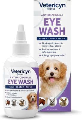
A sterile antimicrobial rinse that flushes irritants, pollen, and discharge out of the eye and helps lift tear stains. A sensible at-home first step for mild irritation, though it does not replace a vet exam for a painful eye.
Webvet may earn a commission when you click through to Chewy, at no extra cost to you.
How does a dog get uveitis?
Ocular causes start in or on the eye:
- Trauma: a scratch, blunt injury, or foreign body that bruises the internal eye.
- Corneal ulceration: a surface ulcer sets off reflex uveitis inside the eye, one of the most common triggers in dogs.
- Lens-induced uveitis: protein leaking from a cataract or a ruptured lens capsule inflames the uvea.
- Cancer inside the eye: primary ocular tumors are less common but important to rule out.
Systemic causes arrive through the bloodstream, which is why canine uveitis is often a red flag for whole-body disease:
- Infections: tick-borne diseases (Ehrlichia, Rocky Mountain spotted fever, anaplasmosis), fungal infections (blastomycosis, histoplasmosis), leptospirosis, and others.
- Immune-mediated disease: the immune system mistakenly attacks eye tissue.
- Cancer elsewhere: lymphoma and other tumors can seed inflammation into the eye.
- High blood pressure or metabolic disease: systemic hypertension and related conditions.
- Idiopathic: in many dogs no cause is found despite a full workup, and the case is labeled idiopathic.
What triggers uveitis flare-ups?
For dogs with an underlying or recurrent condition, flare-ups tend to follow a pattern. Common triggers include:
- A new eye injury or corneal scratch that re-ignites inflammation.
- A relapse of an underlying systemic disease, such as a tick-borne infection.
- Stopping anti-inflammatory eye medication too early, before the eye has fully settled.
- Progression of a cataract, which leaks lens protein into the eye over time.
- Immune-mediated conditions cycling on their own, especially in predisposed breeds.
Because a flare can look identical to the first episode, any return of redness, squinting, or cloudiness deserves a prompt recheck rather than a repeat of leftover drops.
Lens-Induced and Cataract-Related Uveitis
Lens induced uveitis in dogs deserves more than a line on a causes list. It is common, frequently missed, and impossible to fix by treating the eye alone. The lens sits behind the iris, walled off from the immune system. When that isolation breaks down, the body treats lens protein as foreign.
- Phacolytic uveitis: a maturing or hypermature cataract leaks small amounts of lens protein through an intact capsule. The result is low-grade, grumbling inflammation that comes and goes for months.
- Phacoclastic uveitis: the lens capsule actually ruptures, usually from trauma or a rapidly swelling diabetic cataract. Protein floods the eye at once and the inflammation is severe, painful, and sight-threatening within days.
Cataract induced uveitis in dogs is especially common in diabetics. Diabetic cataracts can form in weeks rather than years, and that speed both swells the lens and stresses the capsule. A newly blind diabetic dog whose eyes then turn red is a classic presentation, and the diabetes has to be managed alongside the eye.
This also explains why suppressing the inflammation without addressing the lens tends to fail. The protein keeps leaking, so the drops are holding back a source that never stops. Ophthalmologists routinely control lens-induced inflammation first, then evaluate whether removing the cataract is the durable answer.
When Uveitis Signals Cancer, Lymphoma, or Diabetes
This is the section that explains why your veterinarian wants bloodwork for what looks like an eye problem. The uvea is one of the most blood-rich tissues in the body, so disease circulating anywhere can announce itself there first. Sometimes the eye is the earliest visible sign of a serious systemic illness.
Canine uveitis and cancer are linked closely enough that oncology belongs in the differential for an older dog with no eye injury. Lymphoma is the tumor most likely to appear in the eye, and uveitis in dogs with lymphoma is often bilateral, because the disease arrives through the bloodstream.
- Both eyes affected at the same time, with no history of injury.
- Unexplained weight loss or a drop in appetite over recent weeks.
- Enlarged lymph nodes under the jaw, in front of the shoulders, or behind the knees.
- Drinking and urinating noticeably more than usual.
- Lethargy, exercise intolerance, or new breathing changes.
- Pale gums, easy bruising, or nosebleeds, which can point to a clotting or blood-pressure problem.
Uveitis in dogs with diabetes usually travels the lens-induced route described above, driven by fast-forming diabetic cataracts. It can also involve poor healing and higher infection risk. Either way, blood glucose control is part of the eye treatment plan, not a separate project.
None of this means a red eye is likely to be cancer. Most cases are not. It means that wait-and-see is the wrong instinct here, because the small number of cases that are serious are exactly the ones where early detection changes the outcome.
 Sponsored
Vet care that's always there
Unlimited video calls and messaging with licensed vets for less than a typical vet visit. One membership covers up to five pets.
Use code Pets60
Plans start at $12/month · covers up to 5 pets
- Licensed vets in most states
- Rx for 150+ conditions
- 4.7 on Trustpilot
Is Your Dog's Breed at Higher Risk? (Golden Retriever Uveitis & Other Predispositions)
Most consumer articles skip this entirely, but breed and age genuinely shift the odds. Some dogs carry an inherited tendency toward inflammatory eye disease.


The best-documented example is golden retriever uveitis (often abbreviated GRU), also called golden retriever pigmentary uveitis. It is a distinct, inherited syndrome recognized by veterinary ophthalmologists in which affected Goldens, typically middle-aged and older, develop chronic low-grade inflammation, pigment dispersal onto the lens, and fluid-filled cysts in the eye. GRU is important because it is often bilateral, progressive, and a leading cause of secondary glaucoma in the breed, so at-risk Goldens benefit from routine ophthalmology screening even before symptoms appear.
| Breed / Group | Risk Note |
|---|---|
| Golden Retriever | Inherited golden retriever uveitis (GRU / pigmentary uveitis); screen from middle age |
| Great Dane, German Shepherd | Reported predisposition to uveitis and related inflammatory eye disease |
| Northern breeds (Akita, Samoyed, Siberian Husky) | Higher risk of uveodermatologic (VKH-like) immune disease affecting the eyes |
| Any breed with a cataract | Lens-induced uveitis risk rises as the cataract matures |
| Middle-aged to senior dogs | Systemic causes (infection, cancer, hypertension) become more likely with age |
Age matters too. A young dog with uveitis is more likely to have an infectious or trauma-related cause, while a senior dog raises the index of suspicion for cancer, high blood pressure, or a maturing cataract. None of this replaces a diagnostic workup, but it helps your veterinarian prioritize the search for a cause.
Pigmentary Uveitis in Golden Retrievers (GRU): What Owners Should Know
Pigmentary uveitis in dogs is essentially a Golden Retriever disease. Formally called golden retriever pigmentary uveitis, it is an inherited syndrome ophthalmologists have tracked for decades, and the clearest example of breed predisposition in canine eye disease. Almost no consumer article names it, so many Golden owners meet it late.
GRU is not the same as a one-off inflamed eye. It is a slow, low-grade, chronic process that quietly damages the drainage structures of the eye over years. Onset is typically middle-aged to senior, most often between roughly 8 and 11 years, and it is usually bilateral even when one eye looks worse.
- Radial pigment lines: fine strands of pigment deposited on the front surface of the lens capsule, arranged like spokes. This is the signature finding.
- Uveal cysts: thin-walled, fluid-filled cysts floating in the front chamber or attached behind the iris.
- Persistent faint aqueous flare, meaning low-grade inflammation is present even when the eye looks calm.
- A slow shift in how the eye reflects light, or a subtle haze that owners notice in photographs before they notice it in person.
- Rising intraocular pressure over time as pigment and inflammatory debris clog the drainage angle.
- Cataract formation in the affected eye as chronic inflammation takes its toll.
Pigmentary uveitis in dogs treatment is about control rather than cure. Ophthalmologists typically prescribe long-term topical anti-inflammatory drops at the lowest effective frequency, then monitor pressure on a schedule. The single most important goal is catching secondary glaucoma early, because glaucoma, not the uveitis itself, is what blinds most affected Goldens.
- Expect lifelong medication rather than a finite course, with the dose adjusted at rechecks.
- Pressure checks every few months are the core of monitoring, even when the eye looks fine.
- Report any new redness, squinting, or cloudiness between visits instead of waiting for the next appointment.
- Ask whether both eyes should be treated, since GRU is usually bilateral even when only one eye is symptomatic.
Because GRU is inherited, it also matters for breeding decisions. Affected dogs should not be bred, and screening results are worth asking about when you buy a Golden Retriever puppy. Responsible breeders track eye exam status across their lines. Reference standards are published by the American College of Veterinary Ophthalmologists.
How Vets Diagnose Uveitis in Dogs
Diagnosis has two jobs: confirm that the eye is truly inflamed inside, and hunt for the underlying cause driving it.

Your veterinarian will typically work through these steps:
- Full ophthalmic exam: checking the eye with magnification and a bright light, often a slit lamp, to look for inflammatory cells, flare, and iris changes.
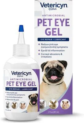
A lubricating antimicrobial gel that clings to the eye surface longer than a rinse, easing conjunctivitis symptoms, lid inflammation, and irritation from minor corneal abrasions. For dogs, cats, and small pets.
Webvet may earn a commission when you click through to Chewy, at no extra cost to you.
- Intraocular pressure (IOP) test: a quick, painless tonometer reading. Uveitis usually lowers pressure, while a rising pressure warns of secondary glaucoma.
- Fluorescein stain: an orange dye that reveals a corneal ulcer, which changes the safe treatment plan (some steroids are unsafe over an ulcer).
- Bloodwork and infectious-disease panel: to screen for tick-borne infection, fungal disease, organ problems, and cancer, especially when both eyes are affected.
- Blood pressure check and imaging: chest or abdominal imaging may be added when a systemic cause is suspected.
- Referral to a veterinary ophthalmologist: recommended for severe, recurrent, or diagnostically puzzling cases, and for breeds like Goldens with suspected GRU.
Because uveitis so often reflects whole-body disease, this systemic search is not over-testing, it is the point. Finding the cause is what lets treatment actually work instead of just masking signs. Some ophthalmologists also recommend a vet-formulated antioxidant such as Ocu-GLO Vision Supplement as ongoing nutritional support for dogs with inflammatory eye disease, introduced alongside (never instead of) the prescribed medical plan.
What Actually Happens at the Visit
Knowing the sequence removes a lot of anxiety, and it explains why the invoice looks the way it does. Most uveitis appointments follow the same path, and each step narrows the possibilities before the next one is added.
- History and observation: when the change started, whether one eye or both, any trauma, current medications, and any systemic signs such as thirst or weight loss.
- Tonometry: a tonometer taps the eye surface painlessly to read intraocular pressure. Uveitis typically drives pressure low, often below the normal range, so a low reading supports the diagnosis. A high reading in an inflamed eye is a red flag for secondary glaucoma.
- Fluorescein stain: orange dye is dropped on the eye and viewed under blue light. It lights up any corneal ulcer, and this result decides whether steroid drops are safe to use at all.
- Magnified internal exam: a slit lamp reveals inflammatory cells, aqueous flare, pigment changes, cysts, and whether the iris is sticking to the lens.
- Retinal exam: the pupil may be dilated to look at the back of the eye for chorioretinitis, hemorrhage, or detachment.
- Bloodwork and infectious-disease panels: added when there is no obvious eye injury, when both eyes are affected, or when the dog is systemically unwell.
- Blood pressure and imaging: chest radiographs or abdominal ultrasound follow when cancer or systemic disease is suspected.
- Ophthalmologist referral: recommended for severe, recurrent, bilateral, or diagnostically unclear cases, and for Goldens with suspected pigmentary uveitis.
Ask for the actual pressure numbers and write them down. They are the single most useful thing to carry from visit to visit, because the trend across rechecks tells you and your veterinarian whether treatment is working long before the eye looks fully normal.
Treatment Options for Uveitis in Dogs
The goal of uveitis in dogs treatment is twofold: calm the inflammation fast to protect the eye, and treat the underlying cause so it does not simply return. Most cases are managed medically, and many dogs are treated at home with a strict eye-drop schedule.

What is the fastest way to cure uveitis?
There is no instant cure, and the fastest real path to resolution is early veterinary diagnosis plus aggressive control of both the inflammation and its cause. The core of anterior uveitis dog care usually combines several medication classes:
| Medication Class | Purpose | Typical Use |
|---|---|---|
| Topical corticosteroid (e.g. prednisolone acetate) | Powerful anti-inflammatory inside the eye | First-line, but only when no corneal ulcer is present |
| Topical NSAID (e.g. diclofenac, ketorolac) | Reduces inflammation without steroid risks | Used when steroids are unsafe (active ulcer) or added on |
| Mydriatic / cycloplegic (atropine) | Dilates the pupil, eases painful spasm, prevents scarring | Comfort and to stop the iris sticking down |
| Systemic anti-inflammatory or antibiotic/antifungal | Treats body-wide inflammation or the underlying infection | Oral meds chosen to match the identified cause |
Prescription dog uveitis eye drops do the heavy lifting, and the schedule can be demanding (sometimes every few hours at first). Between medicated doses, a lubricating gel such as Optixcare Eye Lube Plus can keep the eye surface comfortable, but it is an adjunct only and never replaces the anti-inflammatory drops your vet prescribes. The single biggest mistake owners make is stopping drops early once the eye looks better, which lets inflammation rebound. Always taper on your veterinarian's schedule and return for the pressure recheck.

A sterile lubricating gel that soothes and moisturizes dry, irritated eyes and helps support the tear film in dogs and cats prone to dryness. A gentle, vet-shelf staple for everyday eye comfort.
Webvet may earn a commission when you click through to Chewy, at no extra cost to you.
Eye Drops for Uveitis in Dogs: What Each One Actually Does
Owners are usually sent home with two or three small bottles and very little explanation of why each exists. Knowing the job of each drop makes the schedule easier to keep and makes it obvious why skipping one is not a neutral decision.
- Topical corticosteroid drops shut down inflammation inside the eye. They are the most powerful option and the reason vision is often saved, but they are withheld until a fluorescein stain confirms there is no corneal ulcer, because steroids can worsen an ulcer badly.
- Topical NSAID drops reduce inflammation without steroid risk. They are the choice when an ulcer is present, and are sometimes layered with a steroid in stubborn cases.
- Atropine, a mydriatic and cycloplegic drop, dilates the pupil and paralyzes the painful iris spasm. It is a pain-control drug as much as anything, and it stops the iris scarring down onto the lens.
- Oral anti-inflammatories or immune-modulating drugs treat inflammation the drops cannot reach, particularly in posterior uveitis and panuveitis.
- Targeted therapy for the underlying cause: antibiotics for a tick-borne infection, antifungals for systemic fungal disease, insulin adjustment for a diabetic, oncology care where cancer is found.
- Lubricating drops for surface comfort, used between medicated doses and never in place of them.
Order matters when several drops are due at once. Wait about five minutes between different medications so the first is not washed straight out by the second, and give ointments last. Your veterinary team will tell you the order they want; ask for it in writing.
How to Give Eye Drops to a Painful Eye
A sore eye makes a normally cooperative dog head-shy fast. The goal is a calm, quick, repeatable routine that does not turn medication time into a wrestling match, because a dog that learns to hide will cost you doses over a multi-week course.
- Warm the bottle briefly in your hand. Cold drops startle dogs more than the drop itself does.
- Sit beside your dog rather than looming over the face, and steady the head from underneath the chin with your non-dominant hand.
- Approach from behind and above the head so the bottle never comes at the eye from the front, which most dogs flinch from.
- Tilt the nose slightly upward, use your thumb to lift the upper lid, and let the drop fall into the eye without the tip ever touching the surface.
- Let your dog blink naturally rather than holding the lids shut. Blinking spreads the medication.
- Follow immediately with a treat every single time, so the routine builds a positive association instead of dread.
- If your dog resists hard or the eye looks worse after dosing, stop and call the clinic rather than forcing it.
How Much Does Uveitis Treatment Cost?
How much does it cost to treat uveitis in dogs? It is the question every owner has and almost no article answers. Real numbers vary by region, cause, and severity, but here is a realistic framework so you can plan. These are typical ranges, not quotes, and your clinic can give an exact estimate.
| Cost Tier | Typical Range | What's Included |
|---|---|---|
| General-practice vet visit | $60 to $250 | Exam, IOP check, fluorescein stain, initial drops |
| Diagnostic workup for cause | $150 to $600+ | Bloodwork, tick/fungal panels, blood pressure, sometimes imaging |
| Emergency (ER) visit | $150 to $500+ | After-hours exam and stabilization for a painful or acute eye |
| Veterinary ophthalmologist referral | $250 to $600+ per visit | Specialist exam, advanced testing, tailored treatment plan |
| Ongoing medications | $30 to $150+ per month | Prescription eye drops and any oral systemic therapy |
For a straightforward reflex-uveitis case from a healing ulcer, total costs often land in the low hundreds. A recurrent or systemic case that needs specialist care, repeated panels, and long-term medication can run higher over time. This is exactly the scenario pet insurance is designed for, since eye emergencies are common and treatment is ongoing.
Insurance, Long-Term Drops, and the Worst-Case Number
The one-visit price is only part of the picture. What determines the true cost to treat uveitis in dogs is whether the case resolves or becomes chronic, and whether it stays with your regular vet or moves to a specialist. Here is the honest longer view.
- Ophthalmologist referral consults typically run higher than a general-practice visit and often include advanced imaging, but they frequently shorten the total course by getting the diagnosis right the first time.
- Long-term maintenance drops for a chronic or GRU case commonly land in the range of a modest monthly medication bill, sustained for years rather than weeks.
- Recheck pressure exams every few months add a recurring line item that is easy to underestimate at the start.
- Enucleation, surgical removal of a blind and painful eye, is the worst-case comparator and typically costs many times a single early exam.
- Repeat emergency visits for flare-ups are usually the most expensive way to manage a condition that regular rechecks would have caught earlier.
On insurance, the timing rule is unforgiving. Uveitis diagnosed before a policy starts, or during a waiting period, is treated as pre-existing and generally excluded, and in a breed like the Golden Retriever an insurer may exclude the whole eye. Enroll before symptoms appear or the coverage will not be there.
Home Care: What You Can (and Can't) Do at Home
Owners searching for uveitis in dogs home remedies usually want to help while they arrange care, and that instinct is good, but the honest answer is that there is no home remedy that treats uveitis. The inflammation is inside the eye and requires prescription medication. What you can do at home is supportive and comfort-focused.

For simple hygiene around the eye during a medicated course, a dog-specific saline product such as Nutri-Vet Dog Eye Rinse & Drops can wipe away crust and discharge without stinging. Keep it strictly separate from the medicated drops and never let it replace them. Our guides to how to clean dog eyes and the best dog eye drops for general comfort cover safe day-to-day hygiene, while dog eye discharge and watery eyes help you tell a supportive-care symptom from a red flag.
Natural Treatment for Uveitis in Dogs: The Honest Answer
Searches for natural treatment for uveitis in dogs and for uveitis in dogs treatment at home run high, and the intent behind them is good. The answer has to be direct: nothing available over the counter or in a kitchen reduces inflammation inside the eye, and delay is the thing that costs vision.
- Chamomile, green tea, or herbal eye washes: soothing on skin, but they do not cross into the eye's interior and can introduce contaminants to an already inflamed surface.
- Colloidal silver: no evidence of benefit for intraocular inflammation and a real risk of irritation.
- Saline or boric acid eye rinses: fine for wiping crust from the lids, but they treat nothing inside the eye.
- Turmeric, omega-3, or antioxidant supplements: reasonable general-health additions if your vet approves them, and sometimes recommended as long-term nutritional support, but they are not a treatment for an active flare.
- Human antihistamines or redness-relief drops: these constrict surface vessels and can mask the redness that tells your vet how bad the inflammation is, delaying the real diagnosis.
- Warm or cold compresses: comfort measures only, and pressure on an eye with possible bleeding or high pressure can do harm.
What is genuinely safe to do at home is supportive, and it matters more than owners expect. It keeps your dog comfortable and stops self-inflicted damage while the prescribed medication does the actual work.
- Keep the e-collar on continuously, including overnight. One good rub against furniture can turn inflammation into a corneal ulcer.
- Dim the lights and close blinds, since photophobia makes bright rooms genuinely painful.
- Set phone alarms for every dose, because uveitis drop schedules are frequent and easy to lose track of.
- Wipe discharge away from the outer lids with a clean, damp cloth, working from the inner corner outward.
- Skip walks in bright midday sun and swap in shaded or evening outings during the acute phase.
- Keep other pets from roughhousing with your dog until the eye is cleared at recheck.
Uveitis vs. Other Dog Eye Conditions
A red or cloudy eye has several possible explanations, and owners understandably struggle to tell them apart. This quick-reference table helps you self-triage before the visit. It does not replace an exam, and any painful eye still warrants a same-day call.
| Condition | Redness | Discharge | Pain Level | Urgency |
|---|---|---|---|---|
| Uveitis | Deep, whole-eye redness | Watery, light-sensitive | High (aching, internal) | Same-day vet |
| [Conjunctivitis (pink eye)](https://www.webvet.com/dog-conjunctivitis-pink-eye/) | Surface redness of the lining | Often thick or gooey | Mild to moderate | Prompt, rarely emergent |
| [Corneal ulcer](https://www.webvet.com/corneal-ulcer-in-dogs/) | Redness plus squinting | Watery to mucoid | High, very light-sensitive | Same-day vet |
| [Glaucoma](https://www.webvet.com/glaucoma-in-dogs/) | Red, often a bulging eye | Minimal | Severe | Emergency |
| [Cataracts](https://www.webvet.com/cataracts-in-dogs/) | Usually none | None | None (unless lens-induced uveitis) | Routine to prompt |
Cloudiness is the trickiest overlap. A hazy, painful eye suggests uveitis or glaucoma, while a slowly cloudy, comfortable eye in an older dog is often harmless lens aging rather than disease. Our guide to cloudy eyes in senior dogs walks through that distinction, and if infection is on your mind, see dog eye infection. The key uveitis vs conjunctivitis in dogs takeaway: conjunctivitis irritates the surface lining and is usually less painful, while uveitis inflames the inside of the eye and is a deeper, more urgent problem.
How to Use the Comparison Table Without Self-Diagnosing
Pain level is the most useful column in that table, and it is the one owners tend to skip. A dog with surface irritation blinks and rubs. A dog with internal inflammation squints hard, avoids light, and often goes quiet or off its food. That behavioral gap separates the urgent from the merely annoying.
On uveitis vs conjunctivitis, the practical test is depth. Conjunctivitis (pink eye) in dogs reddens the thin lining around the eye and usually produces thicker discharge with only mild discomfort. Uveitis reddens the eye more deeply, produces watery tearing, and hurts considerably more.
- Thick or gooey discharge with a comfortable dog points toward conjunctivitis rather than uveitis.
- A hard squint with watery tearing and a small pupil points toward uveitis.
- Sudden severe pain with an enlarged, hard-feeling eye points toward glaucoma in dogs and is an emergency.
- A visible surface divot or intense pain after a scratch points toward corneal ulcers in dogs, which can also trigger reflex uveitis at the same time.
- Slow, painless clouding in an older dog is more often lens aging or a cataract than active inflammation.
Settle the dog conjunctivitis vs uveitis question at the clinic, not at home. Conditions can coexist, which is where self-triage runs out. An ulcer plus reflex uveitis is common, and treating only what you can see leaves half of it untreated. Use the table to decide how fast to call.
Prognosis: Will Uveitis in Dogs Go Away?
The uveitis in dogs prognosis is genuinely case-dependent, and that honesty matters more than a falsely reassuring one-liner.
- 1Recovery depends almost entirely on the cause and how quickly treatment started.
- 2Reflex uveitis from a corneal ulcer usually resolves once the ulcer heals.
- 3Infectious cases often clear when the underlying infection is treated.
- 4Immune-mediated, breed-related (GRU), and idiopathic cases tend to need long-term management and can recur.
- 5Untreated or delayed cases carry the highest risk of glaucoma, cataracts, and permanent blindness.
Can dogs recover from uveitis?
Yes, many dogs recover fully, especially when the cause is a treatable one like a healing ulcer or a treatable infection and therapy starts early. Recovery is not always quick: uncomplicated cases may settle over one to a few weeks of drops, while cases tied to systemic disease follow the timeline of that disease. The eye is fragile during inflammation, so completing the full medication course and attending pressure rechecks is what separates a good outcome from a relapse.
Will uveitis in dogs go away?
It can go away, but "gone" is not always the same as "cured for good." When a single cause is found and fixed, the uveitis typically resolves and stays gone. When the cause is chronic (immune-mediated disease, GRU, or unidentifiable idiopathic uveitis), the goal shifts from cure to control, keeping inflammation suppressed and rechecking regularly to catch flares early. The most serious risk of leaving it untreated is secondary glaucoma, which can blind the eye and is far harder to reverse than the original inflammation.
Recovery Timeline: What to Expect Week by Week
Can uveitis in dogs be cured? The truthful answer is cause-dependent, and a timeline makes that concrete. What follows is a typical course for an uncomplicated anterior case treated promptly. Complicated, bilateral, or systemic cases run longer and are guided entirely by your veterinarian.

| Timeframe | What Typically Improves | Vet Touchpoint | Your Job |
|---|---|---|---|
| First 24 to 72 hours | Pain eases as the pupil dilates; squinting softens | Initial exam, stain, pressure reading | Start every drop on time; e-collar on; dim the room |
| Week 1 | Redness and tearing visibly reduce; appetite returns | First recheck with repeat pressure | Keep the full schedule even as the eye looks better |
| Weeks 2 to 4 | Haze clears; pupil responds normally again | Recheck; taper begins only if inflammation is gone | Never taper on your own judgment |
| Month 2 and beyond | Cause-dependent: resolution or long-term control | Follow-up on the underlying disease | Watch for flare signs; keep recheck appointments |
The taper is where most relapses are made. Drops are reduced in steps, and each step is a small test of whether the eye can hold steady with less medication. Cutting the frequency yourself because the eye looks clear is the single most common reason a settled case flares back within days.
- Pain returns after a period of comfort: renewed squinting, hiding, or reluctance to be touched near the head.
- Redness deepens again or spreads across more of the white of the eye.
- Cloudiness expands rather than shrinking between doses.
- The eye looks larger, feels firmer, or the pupil stops responding to light, all of which suggest pressure is rising.
- Vision seems worse: hesitating on stairs, bumping furniture, or losing tossed treats.
- The second eye starts showing any of the above, which changes the diagnostic picture entirely.
Chronic and Recurrent Uveitis: Long-Term Management
Chronic uveitis in dogs is the reality for immune-mediated cases, golden retriever pigmentary uveitis, and many idiopathic cases where no cause is ever found. The frame shifts from cure to control, and that is not a failure of treatment. Well-controlled chronic uveitis is compatible with a completely normal life.
- A maintenance drop schedule, typically the lowest frequency that keeps the eye quiet, reviewed at every recheck.
- Scheduled pressure monitoring every few months, whether or not anything looks wrong.
- A written flare plan so you know exactly who to call and what to do out of hours.
- Watching for synechiae, where the iris scars to the lens and permanently distorts the pupil.
- Watching for inflammatory cataract formation, which chronic inflammation accelerates.
- Watching for secondary glaucoma, the complication that most often ends in enucleation when it is missed.
On the question owners ask quietly: is uveitis in dogs contagious? No. The inflammation itself cannot pass to other pets or to people. Some underlying infectious causes are a different matter, and a few, including leptospirosis and certain tick-borne agents, have zoonotic potential worth raising with your veterinarian.
If an eye is eventually lost, that outcome is far less bleak than owners fear. Dogs navigate confidently on smell and hearing, adapt within weeks to a stable home layout, and removing a chronically painful blind eye usually returns a dog to a comfort level its owners had forgotten was possible.
When to See a Vet Immediately
Uveitis sits on the emergency end of the eye-problem spectrum. Use this checklist to decide when a red or painful eye cannot wait.
Even without those extreme signs, any persistent redness, squinting, cloudiness, or increased tearing is a same-day veterinary matter. There is no safe home version of this diagnosis, and a quick exam with a pressure check is inexpensive insurance against a much larger problem. When in doubt, call your veterinarian and describe what you see. They would far rather check a mild eye than treat an avoidable blindness.
FAQ
Can dogs recover from uveitis?
A: Yes. Many dogs recover fully when the cause is treatable and therapy starts early, such as reflex uveitis from a healing corneal ulcer or a treatable infection. Recovery may take one to several weeks, and finishing the full medication course plus attending pressure rechecks is essential to prevent relapse. Chronic causes are managed rather than cured.
How does a dog get uveitis?
A: A dog develops uveitis from causes inside the eye (trauma, corneal ulcers, cataract-related lens leakage, or ocular tumors) or from body-wide disease reaching the eye through the bloodstream (tick-borne and fungal infections, immune-mediated disease, cancer, or high blood pressure). In many dogs no cause is found despite testing, and the case is called idiopathic.
What triggers uveitis flare-ups?
A: Flare-ups are often triggered by a new eye injury, relapse of an underlying systemic disease, stopping anti-inflammatory drops too early, a progressing cataract leaking lens protein, or the natural cycling of immune-mediated disease in predisposed breeds. Because a flare looks like the first episode, any returning redness or squinting needs a prompt recheck.
What are the first signs of uveitis?
A: Early signs include redness in the white of the eye, squinting or holding the eye partly closed, a smaller-than-normal pupil, increased tearing, mild cloudiness, and light sensitivity. Dogs may paw at the eye or rub their face. These subtle changes are easy to mistake for minor irritation, so a same-day exam is the safe response.
How painful is uveitis in dogs?
A: Uveitis is usually quite painful because the iris and ciliary body are rich in nerve endings and the inflammation causes aching plus painful pupil spasms. Dogs hide pain well, so squinting, light avoidance, reduced appetite, or low energy may be the only outward signs that your dog is genuinely uncomfortable.
What is the fastest way to cure uveitis in dogs?
A: There is no instant cure. The fastest real path is early veterinary diagnosis with aggressive treatment of both the inflammation and its underlying cause, typically anti-inflammatory eye drops (steroid or NSAID), a pupil-dilating atropine drop for comfort, and medication aimed at any identified systemic disease. Home remedies do not treat the internal inflammation.
Will uveitis in dogs go away?
A: It can. When a single cause is found and corrected, uveitis usually resolves and stays gone. Chronic causes such as immune-mediated disease, golden retriever uveitis, or idiopathic cases shift the goal to long-term control rather than cure. Leaving it untreated risks secondary glaucoma and permanent blindness, so early care is critical.
Is uveitis in dogs contagious?
No. Uveitis is inflammation inside your dog's own eye and cannot spread to other pets or to people. Some underlying causes are infectious, however, including tick-borne diseases, fungal infections, and leptospirosis. If a workup finds an infectious cause, ask your veterinarian whether other animals in the household should be tested.
Is uveitis in dogs contagious to humans?
The uveitis itself is not contagious to humans. A small number of the diseases that can cause it, such as leptospirosis and certain fungal or tick-borne infections, do carry zoonotic risk. Basic hygiene and following your veterinarian's guidance is enough in almost every case, but raise the question directly if an infectious cause is identified.
How do vets diagnose uveitis in dogs?
Diagnosis combines a magnified ophthalmic exam for inflammatory cells and aqueous flare, a tonometry reading of intraocular pressure (usually low in uveitis), and a fluorescein stain to check for a corneal ulcer before steroids are used. Bloodwork, infectious-disease panels, blood pressure, and imaging are added when the cause may be systemic.
What is anterior uveitis in dogs?
Anterior uveitis is inflammation of the iris and ciliary body at the front of the eye, and it is by far the most common form in dogs. Signs include deep redness, a constricted pupil, squinting, light sensitivity, and sometimes hyphema (blood) or a hazy front chamber. It requires a same-day veterinary exam.
Is pigmentary uveitis in golden retrievers curable?
No, golden retriever pigmentary uveitis is an inherited, progressive condition that is managed rather than cured. Long-term anti-inflammatory drops and regular intraocular pressure checks keep it controlled and are aimed at preventing secondary glaucoma, which is what most often causes vision loss. Annual ophthalmologist screening from around age 5 is strongly recommended.
Sources

Editor
The Webvet Editorial Team is the in-house group of pet-care editors and writers behind Webvet, operated by Smart Pet Collective. The team researches, writes, and maintains Webvet's pet health, behavior, and medication content. Every article follows a defined editorial process: research from reputable veterinary and scientific sources, careful drafting, mandatory review of medical content by a credentialed veterinarian, and dated publication. Health and medication articles are medically reviewed by a licensed veterinary professional before they go live and are kept current over time.
Veterinarian · BVMS MRCVS
Dr. Pippa Elliott, BVMS, MRCVS, is a veterinarian with nearly 30 years of experience in companion animal practice. Dr. Elliott earned her Bachelor of Veterinary Medicine and Surgery from the University of Glasgow. She was also designated a Member of the Royal College of Veterinary Surgeons. Married with 2 grown-up kids, Dr. Elliott has a naughty Puggle named Poggle, 3 cats and a bearded dragon.


