Wobbler Syndrome in Dogs: Signs, Diagnosis & Treatment
Wobbler syndrome can cause an unsteady gait, weakness, and neck pain. Learn which changes need urgent care and how specialists evaluate treatment and prognosis.

BVMS MRCVS
Medically reviewed by Dr. Pippa Elliott, BVMS MRCVS · Last reviewed

This article contains affiliate links. Webvet may earn a commission when you buy through them, at no extra cost to you.
Wobbler syndrome in dogs is a group of disorders in which structures in the neck compress the cervical spinal cord and sometimes nearby nerve roots. Veterinarians also call it cervical spondylomyelopathy. The condition can produce a swaying or uncoordinated walk, weakness, worn nails, neck pain, and difficulty rising, but no gait pattern seen at home can confirm the diagnosis.
A dog that suddenly cannot stand, is rapidly losing function, has severe neck pain, or is having trouble breathing needs emergency veterinary care. More gradual changes still warrant a prompt examination because orthopedic disease, intervertebral disc disease, vestibular disease, toxins, and other neurologic problems can look similar. A veterinary neurologist may recommend advanced imaging before discussing medical management, surgery, and a realistic prognosis.
 Sponsored
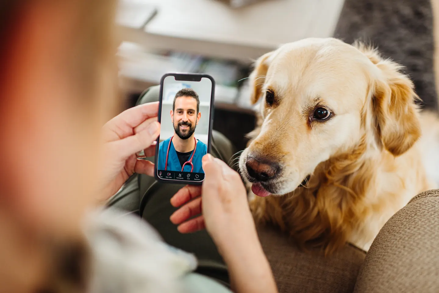
Have a health question about your dog?
Talk to a licensed vet at Dutch over video or message, including nights and weekends.
Use code Pets60
Plans start at $12/month · covers up to 5 pets
- Licensed vets in most states
- Rx for 150+ conditions
- 4.7 on Trustpilot
What Wobbler syndrome is
Cervical spondylomyelopathy describes spinal cord and nerve root compression within the neck. The word "wobbler" refers to the unsteady gait that many affected dogs develop, not to a single cause or a diagnosis that an owner can make by watching a video. Compression may be associated with malformed or poorly aligned vertebrae, changes in joints and supporting tissues, protruding discs, or a combination of static and movement-dependent abnormalities. The location, severity, and duration of compression help determine what a dog feels and how well the limbs work.
The cervical spinal cord carries signals between the brain and all four limbs. When those pathways are disrupted, the hind limbs often show a long, floating, or crossing stride while the front limbs may take short, stiff steps. Some dogs mainly show weakness or scuffed nails. Others hold the head low or resist moving the neck because a disc or nerve root is painful. These differences are one reason the diagnostic process must localize the problem before a treatment choice is made.
- 1Wobbler syndrome in dogs is cervical spinal cord compression, not a descriptive gait diagnosis.
- 2An unsteady walk has many possible causes, so a veterinary assessment with neurologic and orthopedic examination is the first decision point.
- 3Rapid loss of mobility, severe pain, or breathing difficulty should be treated as an emergency.
- 4MRI, CT, or CT myelography may be needed to define the lesion and plan care.
The Ohio State University Wobbler Syndrome Research Program describes the condition as complex and emphasizes that both large and giant breeds can be affected through different patterns of cervical disease. That complexity matters to owners. A diagnosis should identify which spinal levels are involved, whether compression changes with neck position, whether pain is present, and whether another disease better explains the signs.
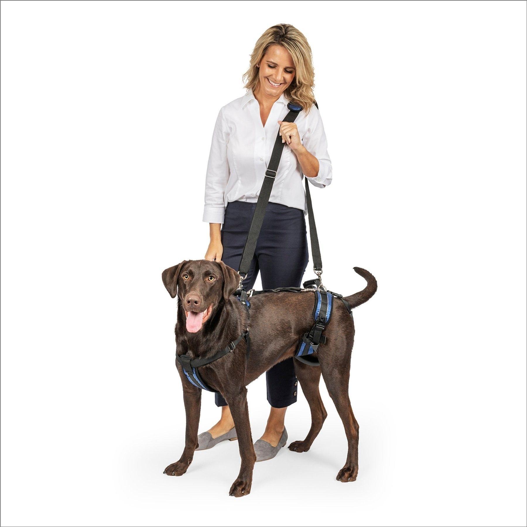
A veterinarian-approved support harness may help caregivers steady a large dog during controlled toilet trips without using the neck as a handle.
Webvet may earn a commission when you click through to Chewy, at no extra cost to you.
Gait changes, neck pain, and emergencies
Early changes can be easy to dismiss on slippery floors or during fast turns. A dog may sway through the hips, place a rear foot too far across the body, drag the tops of the toes, stumble on curbs, or hesitate before stairs. Veterinarians call this incoordination ataxia. Nail wear may be uneven. Some dogs appear clumsy only when walking slowly because deliberate paw placement exposes a proprioceptive deficit. A short safe video can support, but never replace, an examination.
- Changes that deserve a prompt appointment include repeated stumbling, crossing the hind feet, scuffed nails, weakness after ordinary walks, reluctance to raise or lower the head, and a new short-strided front gait.
- Pain may appear as a low head carriage, yelping when turning, reluctance to eat from the floor, guarding the neck, or avoiding play. Not every affected dog is obviously painful.
- A dog that looks dizzy, has a persistent head tilt, circles, or has abnormal eye movements may have vestibular disease rather than cervical spinal cord compression.
- A dog whose back legs tremble may be weak, painful, cold, anxious, or affected by another neurologic or orthopedic problem. The symptom is not specific to Wobbler syndrome.

To tell if your dog has wobblers, watch stride length and rear-limb coordination on a nonslip, level surface, never pulling the neck into a painful range. A short front stride and swaying rear stride raise suspicion.
Many dogs with wobblers are in pain. Ohio State University Wobbler program describes nerve compression as very uncomfortable, often shown by a low head carriage. In a 2023 MRI study of 60 dogs, 55% of dogs in each compression group showed neck pain when examined. Pain control is part of every treatment plan.
For additional context about a nonspecific symptom, see why a dog's back legs may tremble. That guide cannot determine whether an individual dog has cervical disease, but it can help an owner describe timing, triggers, pain, awareness, and recovery. Those observations improve triage because the urgency depends on the whole pattern, not on the word "wobble" alone.
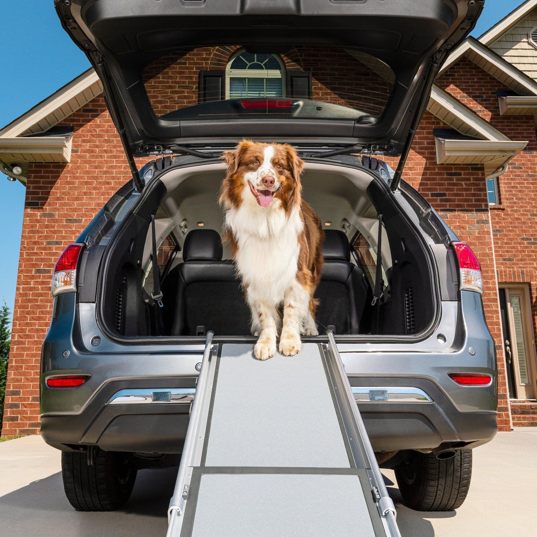
A high-traction ramp may reduce approved jumping during recovery when the veterinarian confirms that controlled ramp use is safe.
Webvet may earn a commission when you click through to Chewy, at no extra cost to you.
Which dogs develop cervical spondylomyelopathy
Great Danes and Doberman Pinschers are the breeds most prone to Wobbler syndrome, although Mastiffs, Rottweilers, Irish Wolfhounds, and other dogs can be affected. The Ohio State University Wobbler program reports a mean age of about 3 years in Great Danes and about 6 years in Dobermans. A breed association changes clinical suspicion, not certainty.
Great Danes more often have vertebral malformation or joint-related compression, while Dobermans more often have disc-associated disease, although these patterns overlap. Most cases occur in large and giant breeds.
Genetics, rapid growth, body size, cervical anatomy, and mechanical forces have all been investigated. Diet has sometimes been blamed, particularly excessive energy or mineral intake during rapid growth, but an owner should not attempt to treat or prevent the condition through an improvised nutrient restriction. Growing large-breed puppies need a complete diet formulated for their life stage and size. Breeders and veterinarians can use family history and careful growth management without promising that either step eliminates risk.
- Breed and age can help prioritize possibilities, but small dogs and mixed-breed dogs still need the same careful localization when signs suggest neck or spinal cord disease.
- A history of trauma does not exclude an underlying developmental or degenerative problem, and an ordinary jump may simply reveal signs that were already developing.
- Neck pain without limb incoordination may come from muscles, joints, discs, infection, inflammation, or nerve roots and should not automatically be labeled Wobblers.
- Related Doberman-specific questions are covered in Wobbler syndrome in Dobermans.
How a veterinarian localizes the problem
The examination begins with a detailed history. The veterinarian will ask when the change began, whether it is steady or episodic, which limbs seem affected first, whether the dog is painful, and whether there was trauma, toxin access, fever, or a medication change. Videos from home can be useful when they show the dog walking naturally, turning in both directions, and rising without being forced. Bring a list of all drugs and supplements because sedation and treatment plans depend on them.
During a neurologic examination, the clinician observes posture, gait, conscious paw placement, spinal reflexes, muscle tone, and cranial nerve function. A pattern of delayed proprioception in all four limbs, often worse behind, with preserved or increased reflexes can localize dysfunction to the cervical spinal cord. Short front steps may result from nerve root discomfort or the dog's attempt to protect its neck. An orthopedic examination is also important because hip, knee, elbow, and shoulder disease can coexist and change the gait.
Localization narrows the list but still does not name the disease. Intervertebral disc disease, vertebral instability, cancer, infection, inflammatory disease, trauma, and congenital abnormalities can affect the same region. A metabolic problem or medication effect may create generalized weakness. If the dog has head tilt, abnormal eye movements, or loss of balance without limb weakness, the clinician may prioritize vestibular causes. If the deficits are confined to the hind limbs, a lesion farther down the spine may be more likely.
- Expect the veterinarian to assess pain without forceful neck movement and to stop if manipulation could worsen injury.
- Basic blood and urine tests do not diagnose Wobbler syndrome, but they can identify anesthesia risks, infection clues, or diseases that mimic weakness.
- Plain radiographs can show vertebral shape, alignment, arthritis, or other bone disease, yet they cannot reliably show the spinal cord or prove the cause of compression.
- Referral is reasonable when deficits are progressing, advanced imaging is needed, pain is difficult to control, or surgery may be considered.
How cervical spondylomyelopathy is diagnosed
MRI is commonly favored because it shows the spinal cord, discs, nerve roots, cerebrospinal fluid spaces, and many soft tissues. It can reveal the location and severity of compression as well as signal changes within the cord that may affect prognosis. The dog usually needs general anesthesia so the neck remains still and safely positioned. The study should be interpreted alongside the neurologic examination because imaging abnormalities can exist in dogs that have few or no clinical signs.
CT provides excellent detail of bone and can help assess vertebral canals, articular processes, and surgical anatomy. CT myelography adds contrast around the spinal cord and may demonstrate compression when MRI is unavailable or when bony detail and dynamic information are priorities. It is invasive and carries different risks, so the neurologist selects the modality for the individual case. Flexed or extended positioning may be considered when dynamic compression is suspected, but deliberately stressing an unstable or severely compressed neck must be carefully controlled.

The Texas A&M Veterinary Medical Teaching Hospital neurology overview lists MRI and CT among the imaging used to diagnose the condition. Owners should ask which levels are compressed, whether the lesion is primarily disc-associated or bone-associated, whether compression is static or dynamic, and whether spinal cord signal changes are present. Those answers are more useful than asking whether a scan looks merely "mild" or "severe."
Testing may also be tailored to competing diagnoses. Cerebrospinal fluid analysis can help when inflammation or infection is suspected, though it is not required for every case. Sampling a suspicious mass may be necessary when cancer is possible. Electrodiagnostic testing can be useful in selected nerve or muscle disorders. A sound workup is therefore a sequence: localize the neurologic problem, rank likely causes, choose tests that can change the plan, and integrate the findings rather than treating an image in isolation.
 Sponsored
Vet care that's always there
Unlimited video calls and messaging with licensed vets for less than a typical vet visit. One membership covers up to five pets.
Use code Pets60
Plans start at $12/month · covers up to 5 pets
- Licensed vets in most states
- Rx for 150+ conditions
- 4.7 on Trustpilot
Medical management and monitored activity
Medical management may be reasonable for dogs with mild or stable deficits, dogs whose imaging and health make surgery less attractive, or families who cannot pursue an operation. The plan can include restricted activity, veterinarian-prescribed pain control or anti-inflammatory medication, weight management, and later rehabilitation. Restriction is not casual rest. It means preventing running, jumping, rough play, stairs, and uncontrolled slipping for the period the clinician specifies, with short supported toilet trips when appropriate.
Medication depends on the source of pain, other diseases, and drugs already being used. Nonsteroidal anti-inflammatory drugs and corticosteroids should not be combined unless a veterinarian has designed a safe transition because the combination increases gastrointestinal and other risks. Gabapentin or other analgesics may be used for selected patients, but sedation can make gait assessment difficult. Never substitute a human pain reliever, adjust a prescription based on internet dosing, or use symptom improvement as permission to resume activity early.
- Ask what level of confinement is required, how long it should last, and which changes should trigger an earlier recheck.
- Use a chest harness rather than pressure on a neck collar when the veterinary team agrees, and keep the leash short enough to prevent lunging.
- Track walking ability, toe scuffing, falls, pain, appetite, urination, bowel movements, and medication effects in a daily log.
- Rehabilitation should begin only after the diagnosis and stability are understood. Exercises that are helpful for one lesion may be unsafe for another.
For a large dog that needs approved assistance during slow toilet trips, a properly fitted full-body support harness can help a caregiver share the load without pulling on the neck. The veterinary or rehabilitation team should demonstrate placement and confirm that straps do not restrict breathing, rub the skin, or let the dog twist. A support device does not make independent walking safe and should never delay reassessment when strength is worsening.
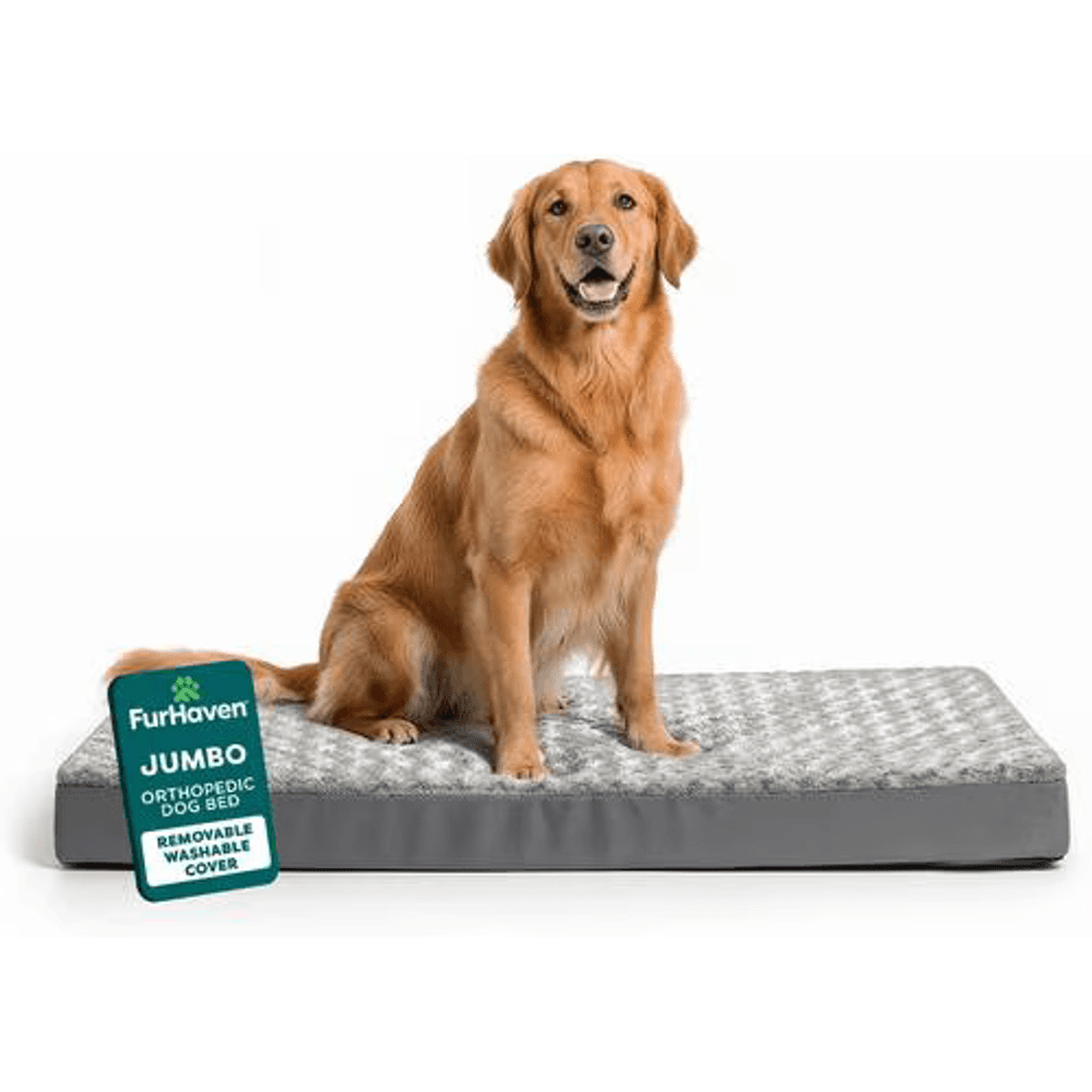
A low, flat orthopedic bed may support comfortable floor-level rest during a veterinarian-directed restriction or recovery plan.
Webvet may earn a commission when you click through to Chewy, at no extra cost to you.
Medical care, surgery, and prognosis
Surgery aims to relieve compression, stabilize an abnormal motion segment, or do both. The technique depends on lesion location, the number of affected levels, whether disease is disc-associated or bone-associated, and whether compression changes with positioning. Procedures may include a ventral slot, distraction and stabilization, or dorsal decompression. Each approach has tradeoffs. Multi-level disease can be difficult, implants can fail, and adjacent spinal segments may develop added stress over time.

The decision is not simply "surgery fixes it" versus "medication avoids risk." A dog with rapid progression, significant compression, and acceptable anesthesia risk may gain more from timely decompression than from repeated conservative trials. A mildly affected, stable dog with extensive multi-level disease or serious medical conditions may reasonably start with medical management. Cost, caregiver capacity, the dog's size, and realistic postoperative restriction also matter because the safest theoretical plan can fail if it cannot be carried out.
| Medical versus surgical factors | May support medical management | May support surgical consultation |
|---|---|---|
| Neurologic status | Mild, stable, independently walking | Progressive weakness, repeated falls, or major functional loss |
| Pain | Controlled with a monitored prescription plan | Persistent or recurrent despite appropriate medication |
| Imaging | Limited compression or anatomy poorly suited to surgery | Defined surgically addressable compression or instability |
| Overall health | Anesthesia or surgical risk is high | Health and recovery capacity are acceptable |
| Home care | Restriction and monitoring are feasible | Postoperative confinement, transport, and rehabilitation are feasible |
| Goal | Maintain comfort and function with surveillance | Relieve compression and preserve or improve function when possible |
| Individual prognosis | Depends on stability, response to rest and medication, and surveillance | Depends on lesion type, number of affected levels, and preoperative walking ability |

Published outcomes are difficult to compare because studies include different breeds, lesions, severities, techniques, and follow-up periods. In a 2023 Journal of Veterinary Internal Medicine MRI study, 40 of 60 dogs (67%) had bone-associated and disc-associated compression at the same site. Mixed lesions like these are one reason an individual prognosis varies.
- Outcome uncertainty is normal, and the questions that influence prognosis are specific: is the dog still walking, how many spinal levels are compressed, and does the spinal cord show signal change?
- Ask about the likelihood of remaining ambulatory, the chance of meaningful improvement, the expected speed of recovery, and the risk of recurrence or adjacent disease.
- Ask what happens next if the chosen plan does not work.
Some dogs improve with either medical or surgical care, some remain stable with limitations, and some progress. Spinal cord damage that has been severe or prolonged may not reverse after decompression. A dog that can still walk before treatment often has a different outlook from one that is nonambulatory, but imaging findings and the specific disease pattern matter. For an individual prognosis, ask the neurologist to explain the best case, most likely case, and worst case for the next weeks, months, and years.
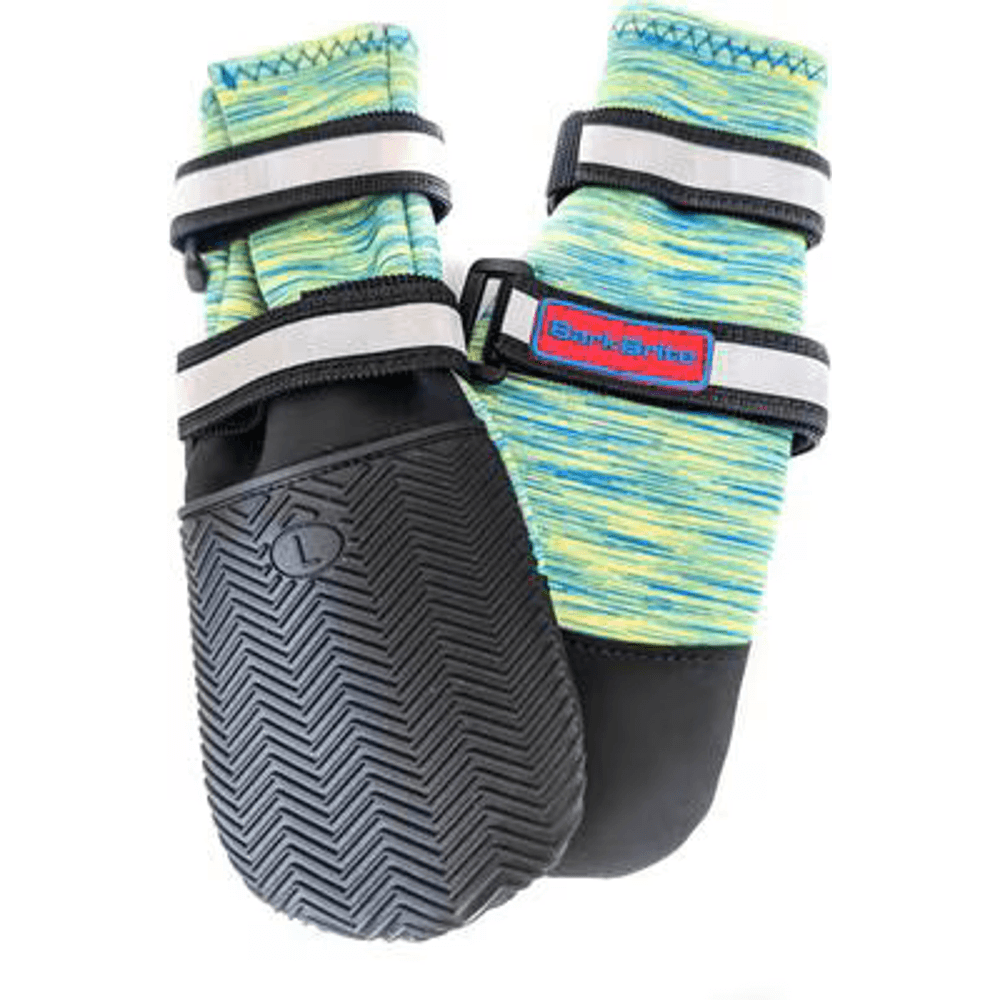
Protective boots may limit paw abrasion during prescribed supported walks, but fit and skin checks should be guided by the veterinary team.
Webvet may earn a commission when you click through to Chewy, at no extra cost to you.
Causes, treatment outcomes, and lifespan
The exact cause of Wobbler syndrome is still not fully known. The Ohio State University Wobbler program describes genetics as a possible factor, but the evidence is not yet clear. Compression usually comes from malformed vertebrae, thickened joints and ligaments, protruding discs, or a mix, and large body size and rapid growth have been studied.
Treatment is either medical management or surgery. The Ohio State University Wobbler program reports that about 50% of medically managed dogs improve, about 30% stay stable, and about 20% worsen, while surgery has a success rate of about 80%. These figures do not predict one dog.
- Medical care means strict activity restriction plus veterinarian-prescribed pain and anti-inflammatory medication.
- Surgery aims to relieve compression or stabilize the affected segment, and the technique depends on the lesion.
- Ohio State reports a mean survival of about 4 years, with similar survival for medical and surgical treatment.
Electroacupuncture is sometimes offered as an add-on. A 2021 literature review of electroacupuncture in dogs with spinal cord injury found too few studies with comparison groups to support firm conclusions, and evidence specific to wobblers is limited. Use it only alongside a neurologist's plan, never in place of it.
How long do dogs live with wobblers?
Many dogs live for years with Wobbler syndrome. The Ohio State University Wobbler program reports a mean survival of about 4 years, similar with medical or surgical treatment. Lifespan depends far more on the individual dog than on the label. Walking ability at diagnosis, pain control, the number of compressed levels, other diseases, and whether a home plan can be sustained all shape the outlook. Repeat neurologic examinations show whether the course is stable or changing.
Building a safer recovery environment
Home management should reduce falls and sudden neck loading while preserving dignity.
- Place nonslip runners between the resting area, door, food, and water.
- Block stairs with a secure gate.
- Keep bowls at the height the veterinary team recommends rather than assuming elevated feeding is always beneficial.
- Separate the dog from rough-playing pets and excited greetings.
- If confinement is prescribed, make the area large enough for comfortable repositioning but too small for pacing or attempts to run.
If the clinician permits access to a low step or vehicle during recovery, a stable high-traction pet ramp may reduce jumping. The slope, width, guard edges, and weight rating must suit a large or weak dog, and the dog should be guided slowly with support. Practice should occur only when the care team says it is safe. A ramp is not appropriate for a dog that cannot control its limbs or for an emergency transport situation requiring immobilization.
A supportive flat orthopedic dog bed can make floor-level rest more comfortable without requiring a dog to climb over high bolsters. Choose a size that lets the dog lie fully extended, a nonslip base, and a washable cover. Check the skin daily, help a weak dog change position as instructed, and keep bedding dry. No mattress prevents spinal cord compression, but a practical rest surface can make a prescribed restriction plan easier to follow.
For dogs that scuff their paws, veterinarian-approved protective dog boots for large paws may reduce abrasion during brief supported outings. Fit and monitoring are essential because a boot can alter proprioceptive feedback, catch on the ground, trap moisture, or create pressure sores. Use protection only for the duration and surfaces the clinician recommends, remove it indoors unless directed otherwise, and inspect toes and pads after every use.
Rehabilitation, rechecks, and measuring progress
Rehabilitation is most useful when it is matched to the lesion, treatment, and phase of healing. Early goals may include safe transfers, maintaining joint range of motion, preventing pressure injuries, and preserving muscle without stressing the neck. Later work may add controlled weight shifting, supported standing, patterned walking, or hydrotherapy. A certified canine rehabilitation professional should coordinate with the neurologist or surgeon because fatigue, pain, and poor limb control can turn an otherwise reasonable exercise into a fall.
Progress should be judged by function over time, not by one good afternoon. Use the same short, safe route and note how much assistance is required, how often paws cross or drag, whether the dog can rise, and how quickly fatigue appears. A weekly video from consistent angles may reveal improvement or decline more clearly than memory. Medication changes must be recorded because sedation or improved pain control can change performance without changing spinal cord compression.
- Call before the scheduled recheck if walking worsens, pain returns, the dog falls more often, or a new limb becomes weak.
- After surgery, report incision redness, swelling, discharge, opening, fever, poor appetite, vomiting, or any neurologic decline promptly.
- Ask when leash walks, stairs, vehicle travel, and independent yard access can resume. These milestones are not interchangeable.
- Continue weight management because excess body weight raises the physical burden of transfers and controlled walking, even though weight loss cannot remove an existing compression.
Quality of life and difficult decisions
Wobbler syndrome does not create one automatic life-expectancy or euthanasia timeline. Quality of life depends on comfort, mobility, continence, appetite, social engagement, sleep, complications, and whether the care plan is sustainable for both dog and caregiver. A dog can have an abnormal gait and still enjoy daily life, while another dog with similar imaging may have uncontrolled pain, fear of falling, or repeated crises. The lived function matters alongside the scan.
A written quality-of-life log reduces the pressure of judging from the worst or best moment. Track comfortable rest, willingness to eat, interest in family, ability to toilet without distress, number of falls, pain episodes, and good versus difficult days. Agree in advance with the veterinarian on thresholds that prompt a medication review, mobility aid, repeat imaging, surgical discussion, or humane euthanasia conversation. Planning those thresholds is responsible care, not giving up.
- When to put a dog down with wobblers is a decision to make with your veterinarian, usually when pain cannot be controlled, the dog can no longer stand or toilet without distress, or falls and crises keep repeating.
- Loss of appetite, withdrawal from family, and more bad days than good days over a sustained period are also important signals.
- A dog with an abnormal gait who is comfortable and engaged may still have a good quality of life.
Caregiver safety also counts. A person should not repeatedly lift a very large dog alone, risk a fall on stairs, or continue a plan that cannot deliver hygiene and pain control. Ask the clinical team for a two-person transfer demonstration, local rehabilitation resources, home-visit options, and realistic costs. If comfort cannot be maintained or the dog experiences persistent distress and loss of meaningful activities despite appropriate care, a veterinarian can help the family evaluate euthanasia compassionately.
Questions to take to the neurology visit
A neurology consultation contains a great deal of information, often while a family is worried. Bring videos, the symptom timeline, prior imaging, laboratory results, medication bottles, and a list of practical home constraints. Ask for the neurologic localization before discussing the scan. Then ask which finding best explains the dog's signs, which abnormalities may be incidental, and which unanswered question could change treatment. This keeps the conversation tied to the patient rather than to a generic Wobbler label.
- Is the compression static, dynamic, or mixed, and how confidently can imaging answer that question?
- Which treatment options are reasonable for this lesion, and what evidence supports each one for a dog with this severity and health profile?
- What deterioration would require emergency care, and whom should we contact after hours?
- What will restriction and rehabilitation look like during the first two weeks, first six weeks, and beyond?
- If we choose medical management now, would delay make later surgery less effective, and what finding would trigger reconsideration?
- How will we define success, and when should the plan be changed if pain or function does not improve?
Before leaving, repeat the plan in your own words and request written instructions. Confirm drug names, doses, timing, activity limits, equipment guidance, recheck timing, and emergency thresholds. Wobbler syndrome is a complex diagnosis, but the next decision should be concrete: protect the dog from falls, control pain safely, complete the tests that will change management, and monitor the functions that matter.
Questions about wobbler syndrome in dogs
These answers cover the questions owners ask most often. Every dog still needs its own veterinary examination. Sudden loss of walking, severe neck pain, or breathing difficulty remains an emergency.
Frequently Asked Questions
How long can a dog live with wobblers?
Many dogs live for years with Wobbler syndrome. The Ohio State University Wobbler program reports a mean survival of about 4 years, similar with medical or surgical treatment. Dogs that are still walking and comfortable when treatment starts often have a better outlook than dogs that cannot stand, and severe or long-standing spinal cord damage may not reverse. Pain control, the number of compressed levels, and the response to the first weeks of treatment are among the most useful guides.
What are the first signs of wobblers?
The first signs often include a swaying hind end, crossing the rear feet, scuffed nails, stumbling on turns, a long floating rear stride, short stiff front steps, or reluctance to move the neck. These findings also occur with orthopedic, vestibular, and other neurologic disorders, so observation alone cannot diagnose the condition.
How fast do wobblers progress?
Wobblers can progress gradually over months or decline suddenly, sometimes after minor trauma. The course depends on the kind of compression, the number of levels involved, activity, and treatment response. Rapidly worsening weakness, repeated falls, loss of walking, severe pain, or breathing difficulty requires emergency care. Keep dated videos between visits.
How to tell if your dog has wobblers?
You cannot confirm Wobbler syndrome at home. Suspicion rises when a large dog has a short, stiff front stride, a wide or crossing rear stride, toe scuffing, or neck pain. A neurologic examination localizes the problem, and MRI or CT is usually needed to confirm compression and rule out look-alike diseases.
Are dogs with wobblers in pain?
Many are. Compression of the spinal cord and nerve roots can cause neck pain, often shown as a low head carriage, yelping when turning, or reluctance to lower the head. In a 2023 MRI study of 60 dogs, neck pain on examination was found in 55% of dogs with combined bone- and disc-associated lesions, so some dogs have weakness without obvious pain. Never give human pain relievers.
When to put a dog down with wobblers?
There is no fixed point. Euthanasia is usually discussed when pain cannot be controlled, the dog can no longer stand or toilet without distress, complications keep recurring, or the care plan cannot be sustained safely. A written quality-of-life log helps the family and veterinarian judge the trend rather than one bad day.
What breeds are prone to Wobblers?
Doberman Pinschers and Great Danes are the most commonly affected breeds, according to the Ohio State University Wobbler program. Mastiffs, Rottweilers, Irish Wolfhounds, and other large or giant breeds are also affected, and small dogs occasionally are. Breed raises suspicion but never makes the diagnosis. See Wobbler syndrome in Dobermans for breed detail.
Sources
- Ohio State University Wobbler Syndrome Research Program
- Texas A&M Veterinary Medical Teaching Hospital: Wobbler Syndrome
- Magnetic resonance imaging and neurologic characterization of combined osseous- and disc-associated cervical spondylomyelopathy in dogs, Journal of Veterinary Internal Medicine (2023)
- Current Aspects Regarding the Clinical Relevance of Electroacupuncture in Dogs with Spinal Cord Injury: A Literature Review, Animals (2021)

Editor
The Webvet Editorial Team is the in-house group of pet-care editors and writers behind Webvet, operated by Smart Pet Collective. The team researches, writes, and maintains Webvet's pet health, behavior, and medication content. Every article follows a defined editorial process: research from reputable veterinary and scientific sources, careful drafting, mandatory review of medical content by a credentialed veterinarian, and dated publication. Health and medication articles are medically reviewed by a licensed veterinary professional before they go live and are kept current over time.
Veterinarian · BVMS MRCVS
Dr. Pippa Elliott, BVMS, MRCVS, is a veterinarian with nearly 30 years of experience in companion animal practice. Dr. Elliott earned her Bachelor of Veterinary Medicine and Surgery from the University of Glasgow. She was also designated a Member of the Royal College of Veterinary Surgeons. Married with 2 grown-up kids, Dr. Elliott has a naughty Puggle named Poggle, 3 cats and a bearded dragon.


