Dog Ear Mite Symptoms: Signs, Pictures and Lookalikes
Recognize possible ear-mite signs, interpret a real parasite micrograph honestly, compare wax and infection lookalikes, and prepare useful observations for a veterinary examination.

BVMS MRCVS
Medically reviewed by Dr. Pippa Elliott, BVMS MRCVS · Last reviewed

This article contains affiliate links. Webvet may earn a commission when you buy through them, at no extra cost to you.
Dog ear mite symptoms can include repeated head shaking, scratching, irritated ears and dark, crumbly debris. Those signs are useful reasons to arrange an examination, but they cannot confirm mites. Yeast, bacterial infection, allergy and foreign material can look similar, and an ear can have more than one problem at once.
The most useful distinction is between seeing an ear change and identifying its cause. A photograph can record what you noticed. An otoscope examination and, when appropriate, a microscopic sample let the veterinarian look for the parasite and other disease. The real microscope photograph below shows an ear mite; it is not a template for diagnosing dark wax at home.
- 1Pictures cannot confirm the cause of dark ear debris.
- 2Record scratching, discharge, odor and whether one or both ears changed.
- 3Mites and a secondary infection can occur together.
- 4Get urgent advice for severe pain, poor balance, a persistent head tilt or a rapidly worsening dog.

 Sponsored
Need flea, tick, or parasite care for your dog?
A licensed vet at Dutch can recommend the right preventive and prescribe it when it's appropriate. Annual members get 20% off their first flea and tick order.
Use code Pets30
Plans start at $11/month · covers up to 5 pets
- Licensed vets in most states
- Rx for 150+ conditions
- 4.7 on Trustpilot
Common dog ear mite symptoms
A dog with ear mites may scratch around the ears, rub its head or shake repeatedly.
The ear canal or flap can look irritated, with dark debris or crusting. Some dogs dislike having the ear touched. These behaviors tell you the ear is uncomfortable; the amount of scratching is not a reliable measure of how many mites are present.
- Repeated head shaking or pawing at the ears.
- Dark waxy or crumbly discharge, sometimes described as coffee grounds.
- Redness, crusts or scratches around the ear opening or flap.
- Hair loss where the dog has been rubbing or scratching.
- A change in how the ear is carried, or reluctance to have the head handled.
The Merck Veterinary Manual notes that itching can vary and an ordinarily upright ear may droop. An odor, wet discharge or pus may indicate a complicated ear problem, including secondary infection. A dog with only a little visible wax can still need assessment, while a heavily waxed ear is not proof of mites. Do not wait for every sign on a checklist.
Puppies, one-sided signs and early changes
Puppies are a useful group to consider because exposure to other young animals may be part of their history. Tell the clinic about littermates, adoption and other household pets. A puppy scratching one ear still needs both ears assessed; what you notice on one side does not establish whether the other is unaffected. Use the same careful description for an adult dog.
There is no reliable owner-facing “early stage” picture that dates an ear-mite infestation. Mild signs can be early, partially treated or caused by something else. A severe-looking ear may include self-trauma or infection. Record when you first noticed a change rather than assigning a stage from an online image. That timeline is more useful at the appointment.
What do ear mites look like?
An actual ear mite, Otodectes cynotis, is a tiny pale arthropod. It is barely visible without magnification and may look like a small moving speck against a dark background.
Most owners notice the ear’s reaction rather than clearly seeing mites. An image of coffee-ground-like discharge records ear debris, not individual parasites; a white spot is not enough to identify one either.

Photograph and attribution source; CC BY-SA 2.0 licence. The original image is reproduced without changes.
This photograph shows the organism’s body and legs under a microscope. It does not show the color, depth or severity of an infected ear canal. Phone cameras, flash, skin pigment and recent cleaning change the appearance of ear photographs, so matching your dog’s ear to a search image cannot choose a medicine. Ear-mite eggs are also not something to diagnose from an ordinary photograph of the flap.
Use pictures to document a change
If your dog is comfortable with handling, an outside view can record swelling, a scratch or discharge at the opening. Take the picture without pushing the camera, a swab or a tool into the canal. Do not hold down a painful dog to get a better image. A short video of head shaking may be more useful than a close-up obtained by force.
- Note the date and which ear is shown.
- Describe the material in plain terms: dark and crumbly, wet, waxy or blood-stained.
- Record any cleaner or medicine used before the photograph.
- Send the image to your veterinary team if they request it; keep the examination appointment.
Pictures of dogs with ear mites often show behavior such as scratching, but that behavior is not specific to parasites. Similarly, a picture of dark material can be an accurate record without being a diagnostic image. The useful question is what has changed for your dog and what needs examining, not which photograph looks most similar.
Ear mites, wax and infection can look alike
Ear mites versus ear wax is not a simple color test. Wax is a normal ear secretion, while an increased amount or a change accompanied by discomfort may reflect disease.
Brown debris or dark gunk can be associated with yeast, bacteria or mites. A dirty-looking ear does not tell you which is present, and washing it clean does not prove the underlying problem is resolved.
| What you notice | What it cannot establish | Useful information for the vet |
|---|---|---|
| Dark crumbs or coffee-ground-like debris | Whether the material contains mites, yeast, bacteria or a mixture. | When it appeared and whether the ear was recently cleaned. |
| Redness, itching or head shaking | Which organism or underlying condition is responsible. | One or both ears, severity and any skin itching elsewhere. |
| A strong smell or wet discharge | A specific bacterial or yeast diagnosis from appearance alone. | Change in odor, discharge, pain and previous medication. |
| A moving pale speck | Reliable parasite identification without a proper examination. | Mention what you saw; avoid trying to extract it yourself. |
The comparison is about the limits of an observation, not permission to ignore it. Persistent dark debris with scratching needs evaluation even if your dog seems otherwise well. Ear mites and yeast can coexist, so a vet may need to treat two problems. A photograph cannot identify the cause or rule out a foreign object deeper in the canal.
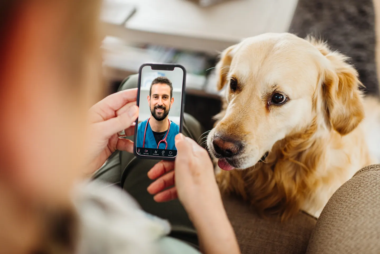 Sponsored
Vet care that's always there
Unlimited video calls and messaging with licensed vets for less than a typical vet visit. One membership covers up to five pets.
Use code Pets30
Plans start at $11/month · covers up to 5 pets
- Licensed vets in most states
- Rx for 150+ conditions
- 4.7 on Trustpilot
How the veterinarian confirms the cause
The veterinarian combines the history with an ear examination. An otoscope provides light and magnification to inspect the canal; a sample examined under a microscope can show mites.
Cytology may also be used to assess yeast, bacteria and inflammation. Ask what was actually found rather than assuming the diagnosis from the label on a bottle.

If the ear is very painful or blocked by debris, the assessment may need pain relief or sedation. VCA’s veterinary guidance explains this examination limit. Do not probe deeper because a first look at home showed nothing. A dog resisting ear handling may be protecting a sore area, not simply being uncooperative.
- When did scratching or discharge first appear?
- Were both ears affected at the same time?
- Has another animal had itchy ears, or joined the household recently?
- Which preventives, drops, wipes or home mixtures have already been used?
- Did a previous treatment help, and for how long?
These details help distinguish a new exposure from recurring disease or incomplete treatment. If you have already cleaned the ear, say when and with what; there is no need to hide it or add more cleaner before the visit. Ask the practice whether they want the ear left untouched so they can assess the original discharge.
When infection is also a possibility
The dog ear infection symptoms guide explains redness, discharge, odor, swelling and clinical photographs in more detail. Those findings overlap with mite-related irritation. An adult dog with repeated ear problems may need investigation for an underlying cause such as allergy, whether or not an earlier episode was blamed on mites.
Do not use infection photographs as a way to rule mites in or out.
The appearance can change after scratching or cleaning, and secondary infection can alter an ear that started with parasites. Tell the veterinarian about skin itching, previous ear episodes and any hearing or balance change so the whole pattern is assessed.
When the symptoms are urgent
Get prompt veterinary advice for severe ear pain, a persistently tilted head, stumbling, unusual eye movements, sudden hearing changes, substantial swelling or a dog that seems generally unwell.
Seizures, collapse or marked loss of balance need urgent assessment. These are not signs to explain away as ordinary ear mites while waiting to see whether drops help.
| Change to report | Next step |
|---|---|
| Itching, debris or odor without marked distress | Arrange an ear examination and ask whether cleaning should wait. |
| Severe pain or sudden ear-flap swelling | Contact the clinic promptly for assessment and safe handling advice. |
| Balance or neurologic change | Seek urgent veterinary advice; do not assume the problem is confined to the outer ear. |
A suddenly ballooned ear flap may be an ear hematoma associated with scratching or head shaking. Do not drain or squeeze it. Ear problems can be painful enough to make an otherwise gentle dog snap, so avoid forceful restraint and let the clinic advise how to transport and handle your dog comfortably.
Why contact with other pets matters
The parasite spreads chiefly through close contact with affected animals, including cats and dogs. A puppy, a newly adopted pet or another animal with itchy ears is relevant history; an indoor dog can still have contact with an affected housemate. For the full transmission, lifecycle and prevention plan, see ear mites in dogs.
When mites are confirmed, the veterinarian should advise care for all exposed household dogs and cats, including those without obvious signs.
Human problems are uncommon, but someone with a new rash or ear symptoms should contact their own clinician. Treating bedding or separating one scratching dog cannot replace an appropriate plan for the animals.
What to do before the appointment
Call the clinic, keep water out of the affected ear and avoid probing the canal. Do not add vinegar, peroxide or oil to test whether the debris disappears. The dog ear mite home-remedy guide explains why household mixtures are not a dependable parasite treatment and what supportive care can safely mean while awaiting assessment.
Ask whether cleaning should wait until the examination. If scratching is injuring the skin, request advice about a suitable protective collar and pain management.
Do not give human pain medicines or use a leftover prescription on your own. Keep product packaging and your brief symptom timeline ready for the visit.
What happens after mites are confirmed
One prescription option is Revolution for dogs, a selamectin product applied to the skin as directed. Manufacturer information identifies its canine ear-mite use and age and health precautions. The linked product is the 40.1–85-pound presentation, not a universal dog dose. The veterinarian must choose the correct weight band; do not put this skin medicine down the ear canal.
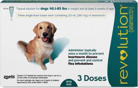
A monthly topical (selamectin) that prevents heartworm and controls fleas, and is well tolerated in herding breeds at the label dose. A prescription pick for dogs who need a non-isoxazoline option.
Webvet may earn a commission when you click through to Chewy, at no extra cost to you.
Bio-Groom Ear Mite Treatment is a different kind of product, applied according to its ear-treatment label only when appropriate. Its manufacturer directions exclude puppies under 12 weeks. A diagnosis and an ear-safety assessment still matter when a product is sold over the counter. Do not treat a painful unexamined ear merely because the package mentions mites.
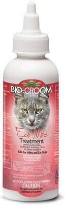
An aloe-enriched medicated treatment that kills ear mites and ear ticks while calming the itch and irritation they cause. For dogs, cats, and rabbits.
Webvet may earn a commission when you click through to Chewy, at no extra cost to you.
Cleaning after the examination
The clinic may recommend cleaning once it is comfortable and compatible with the treatment.
Ask which product to use, how often and whether to leave a gap around medication. An ear that looks cleaner has not necessarily been cleared of mites. If the dog pulls away in pain, stop that attempt and call instead of increasing restraint.
Pet MD Aloe and Eucalyptus Ear Wipes are a possible option for suitable accessible outer-ear debris; they are not parasite treatment and should not be pushed into the canal. Their directions specify dogs over 12 weeks. Keep wipes out of a raw or painful ear unless the veterinary team has specifically approved them.

Webvet may earn a commission when you click through to Chewy, at no extra cost to you.
A cleanser such as Epi-Otic Advanced may fit an individual plan after the ear has been assessed. Cornell’s cleaning guidance explains why cotton swabs inside the canal and irritating household liquids should be avoided. Use the demonstration and directions provided for your dog rather than a generic daily cleaning schedule.
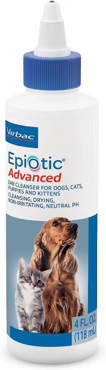
Webvet may earn a commission when you click through to Chewy, at no extra cost to you.
If signs continue after treatment
Keep the scheduled recheck and report whether scratching, odor or discharge has changed.
Persistent symptoms do not automatically mean the product failed to kill mites: inflammation, secondary infection or another original cause may still need attention. Conversely, a quieter dog is not laboratory evidence that parasites are gone. Let the follow-up examination answer that question.
Bring the dates and names of treatments used for every household pet. A missed application, a product used in the wrong place or an untreated contact animal may change the next step. Record what happened without blaming yourself or guessing at a stronger treatment. The useful result is a confirmed explanation and an achievable care plan.
Questions owners ask
Frequently Asked Questions
What can be mistaken for ear mites in dogs?
Wax buildup, yeast or bacterial infection, allergic inflammation and foreign material can produce similar signs. Some of these conditions can coexist with mites. An ear examination and appropriate microscopic testing help distinguish them. Do not choose a mite medicine from discharge color or a picture, especially if previous treatment did not resolve the problem.
Are ear mites painful for dogs?
They can cause substantial irritation, and inflammation, scratching injuries or a secondary infection can add ear pain. A dog that cries, resists handling or cannot settle needs veterinary care. Severe pain or a change in balance deserves prompt assessment rather than repeated cleaning. Ask the clinic how to keep the dog comfortable until the examination.

Editor
The Webvet Editorial Team is the in-house group of pet-care editors and writers behind Webvet, operated by Smart Pet Collective. The team researches, writes, and maintains Webvet's pet health, behavior, and medication content. Every article follows a defined editorial process: research from reputable veterinary and scientific sources, careful drafting, mandatory review of medical content by a credentialed veterinarian, and dated publication. Health and medication articles are medically reviewed by a licensed veterinary professional before they go live and are kept current over time.
Veterinarian · BVMS MRCVS
Dr. Pippa Elliott, BVMS, MRCVS, is a veterinarian with nearly 30 years of experience in companion animal practice. Dr. Elliott earned her Bachelor of Veterinary Medicine and Surgery from the University of Glasgow. She was also designated a Member of the Royal College of Veterinary Surgeons. Married with 2 grown-up kids, Dr. Elliott has a naughty Puggle named Poggle, 3 cats and a bearded dragon.


